Director: Dr. Bryan T. Hackfort
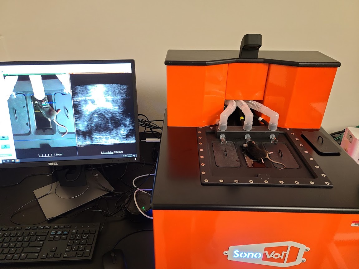
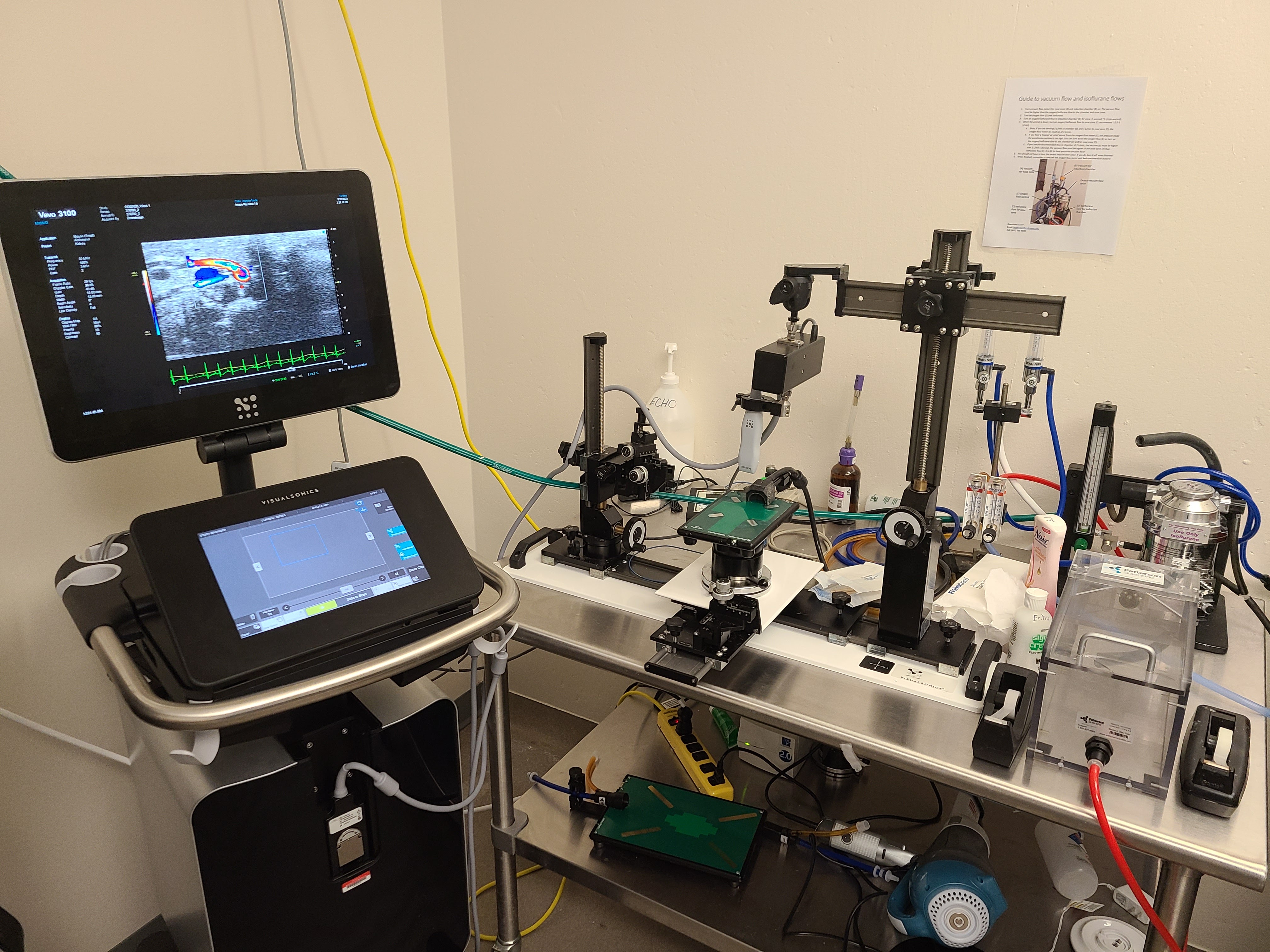
The echocardiography imaging facility consists of an Acuson 512C echocardiograph and associated probes (3-5 MHz and 15 MHz linear array), a Visual Sonics Vevo 3100, a Vevo 3100 with the LAZR-X, and a Perkin Elmer Vega. Each Vevo machine has probes ranging from 15 MHz to 55 MHz. The Vevo systems have injector rails for echo-guided delivery into rodents. Cardiovascular imaging ranges from simple 2D and M-mode indices of cardiac function (EF, FS, dimensions, volumes, wall thickness, etc) to assessment of diastolic function (E/A) and pulsed Doppler flow, 4D imaging, and speckle tracking using Vevo Strain. We also offer longitudinal imaging of tumors with available 3D volumetric analysis. Photoacoustic imaging with the LAZR-X uses a laser in the ranges of 680 nm - 970 nm or 1200 nm – 2000 nm to create a thermoelastic change in chromogens which generates a mechanical signal picked up by ultrasound. This can be used to detect changes in oxygen saturation levels of hemoglobin or other chromogens (ICG, IR-800, etc). Changes in flow can be monitored using non-linear contrast imaging and microbubbles. The Vega system brings high throughput imaging to the Core along with shear wave elastography and acoustic angiography. The core operates on a fee for service basis. Charges range from $60 to $100/hour dependent on technician involvement. Analysis can be done by investigators or by the Core director as an additional charge.