Preclinical Imaging Instrumentation
The Bioimaging Core Facility operates two small bore high field (7 Tesla) Magnetic Resonance Imaging (MRI) and MR Spectroscopy (MRS) systems and one Single Photon Emission Computed Tomography scanner with an integrated X-ray Computed Tomography imaging system (SPECT/CT scanner). In addition, computing equipment is available for image storage, analysis, custom computer program development, and database development.
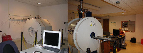
Two animal MRI/MRS systems include a Bruker Biospec MRI and MRS system (7T/21 cm, Bruker, Karlshure, Germany) and a Bruker Pharmascan (7T/16 cm) MRI/MRS system equipped with Resonance Research (Billerica, MA) gradients and shims. The MRI/MRS systems are housed in two adjacent 600 square foot laboratories.
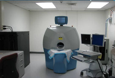
An adjacent laboratory houses the preclinical single photon emission computed tomography (SPECT) and computed tomography (CT) dual scanner (GammaMedica-Ideas (GM-I) Flex Triumph Multimodal Preclinical Platform) equipped with multi pinhole collimators, variable radius of rotation heads (1.5 cm to 17.5 cm) and four interchangeable patient beds to accommodate animals from mice to small primates. GM-I’s new VIVID (Volumetric Image Visualization, Identification and Display) software package has been developed specifically for FLEX Triumph with a streamlined user interface for automated fusion and display, automated loading of SPECT, PET and CT image data, and an automated visualization module for 2D, 3D, and single or multiple (fused) images. The SPECT/CT system is located adjacent to the two MRI laboratories, allowing multimodal imaging of the same animals in both types of scanners.
An adjacent area is subdivided and used for a probe development laboratory and chemical preparation laboratory. The probe laboratory is equipped with a lathe, milling machine, drill press, band saw, PTS frequency synthesizer, 500 MHz digital oscilloscope, transmission and reflection RF sweeper, and basic tools and electronic test equipment to allow machining and development of custom coils and animal holders for projects.
The data analysis laboratory houses three Unix workstations and an array of PC’s for o`ine data analysis. The data analysis laboratory houses 3 Unix workstations and an array of PC’s for offline data analysis. The workstations include one Dell Precision T1700 and two Sun Blade 2500 systems. Database development and data storage is provided by the Research Information Technology Office (RITO) core server.
Computers are loaded with image data analysis programs including Analyze (Biomedical Imaging Resource, Mayo Clinic, Rochester, MN), and Eigentool (Henry Ford Health Sciences, Department of Radiology, Detroit, MI) as well as custom programs for image processing using Unix shell scripts and pipes (Dr. James Ewing, Henry Ford Health Sciences, Department of Neurology, Detroit, MI). Available programs will allow development of efficient processing pipelines tailored to needs of individual projects’ needs. Custom programming methods have also been developed and implemented using Matlab (Mathworks, Inc, Natick, MA) and IDL (Research Systems, Inc., Boulder, CO) on both PC and Sun workstations. Diffusion tensor image analysis and display program developed and supplied by Dr Khader Hasan, University of Texas Medical School, Houston, TX written in IDL. Coregistration of MRI and SPECT has been developed based on custom designed animal holders equipped with MRI and SPECT compatible fiducial markers and geometry compatible with both the SPECT and MRI scanners. Spectroscopic analyses are performed using the LCModel (S. Provencher, Ontario, Canada) spectroscopic fitting package, jMRUI spectroscopic processing and fitting packages in addition to an array of lab-developed image and spectroscopic image processing and analysis programs.