We employed manganese (Mn)-enhanced magnetic resonance imaging (MEMRI) as a noninvasive imaging biomarker to follow disease progression and to assess the abilities of a vasoactive intestinal peptide receptor 2 agonist (LBT-3627) therapies to slow neurodegenerative activities in MPTP intoxicated mice. Notably, LBT-3627-treated, MPTP-intoxicated mice show reduced MEMRI brain signal intensities. These changes paralleled reduced astrogliosis and resulted in sparing of nigral tyrosine hydroxylase neurons. Most importantly, the data suggest that MEMRI can be developed as a biomarker tool to monitor neurotherapeutic responses that are relevant to common neurodegenerative disorders used to improve disease outcomes.
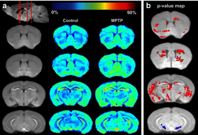 Comparison of MEMRI signal enhancement between controls and MPTP-intoxicated mice at day 2 post-MPTP.
(a) MEMRI enhancement maps. The first column (from left) shows coronal slices of the averaged MEMRI of control mice (n = 5) as an anatomical reference. The second column shows the average enhancement in control mice on the coronal slices. The third column represents the average enhancement of MPTP-intoxicated mice (n = 6). (b) Statistical comparison of MEMRI enhancement between control and MPTP- intoxicated mice. Pixels with a significant increase (p < 0.05) in signal intensity are indicated in red, with decreases indicated in blue.
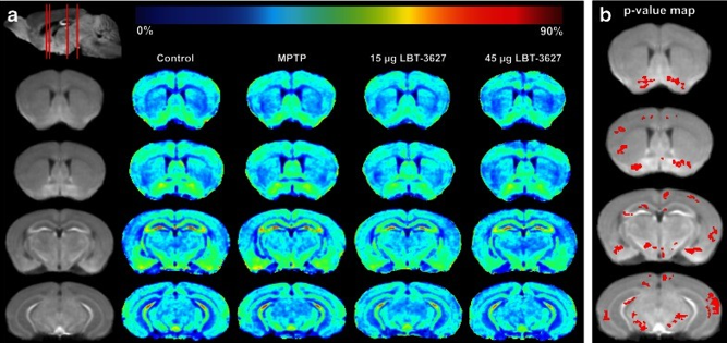
Comparison of MEMRI enhancement between controls and MPTP-intoxicated mice at day 7.
(a) MEMRI enhancement maps. The first column (from left) shows coronal slices of the averaged MEMRI of control mice (n = 5) as an anatomical reference. The sagittal slice (upper left) shows respective coronal positions (red lines). The second column shows the average enhancement in control mice on the coronal slices. The third column represents the average enhancement of MPTP- intoxicated mice (n = 6). The fourth column represents the average enhancement of MPTP-intoxicated mice treated with 15 ug
LBT-3627. The fourth column represents the average enhancement of MPTP-intoxicated mice treated with 45 ug LBT-3627. (b) Statistical comparison of MEMRI enhancement between MPTP-intoxicated mice and MPTP-intoxicated mice treated with 15 ug LBT-3627. It shows the pixels (red color) with significant decrease (p < 0.05) in signal intensity overlaid onto the averaged brain image
For further information, see:
Katherine E. Olson, Aditya N. Bade, Charles R. Schutt, Jingdong Dong, Scott J. Shandler, Michael D. Boska, R. Lee Mosley, Howard E. Gendelman, and Yutong Liu. Manganese-Enhanced Magnetic Resonance Imaging for Detection of Vasoactive Intestinal Peptide Receptor 2 Agonist Therapy in a Model of Parkinson’s Disease. Neurotherapeutics. 2016 Jul; 13(3): 635–646.