- Core Facility Directory
- Clinical Research Resources
- Nebraska Biobank
- Grant Editing Support
- Grant Resource Library
- Centers and Major Programs
- Sponsored Programs Administration (SPA)
- Comparative Medicine
- Government Relations
- Intellectual Property Assistance
- Translational Cores
- Advanced Microscopy Core Facility
- Animal Behavior Core
- Biosafety Level 3 (BSL-3) Core Facility
- Computational Chemistry
- Electron Microscopy
- Epigenomics Core Facility (ECF)
- Flow Cytometry Research Facility
- Genomics Core Facility
- Human Magnetic Resonance Imaging
- Mouse Genome Engineering Core
- Multiomics Mass Spectrometry
- Nanoimaging Core Facility
- READi Core (health informatics)
- Small Animal Imaging
- In Vivo Imaging Core Facility
- Multiphoton Intravital & Tissue Imaging (MITI) Research Core
- MRI/MRS and SPECT/CT Core Facility
- PET Core Facility
- VEVO Ultrasonic Imaging
- Translational Mouse Model Core Facility
- Core Facility Scheduling and Billing
- Backup Freezer Program
The images, 3D reconstructions and digital movies below represent the diverse types of experiments and imaging underway at the MITI core. In addition to intravital imaging of multiple fluorophores in live small rodents, collagen second harmonic imaging, multiphoton imaging of optically cleared samples, and live cell tracking in fresh tissue explants is actively underway.
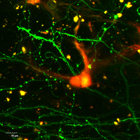
Individual eGFp positive neuron s(green) with clearly delineated dendritic spines are observed in close proximity to a TurboRFP positive astrocyte (red) in the cortex of a live mouse using specialized imaging windows.
Credit: Dr. Padmashri Ragunathan, Dr. Anna Dunaevsky
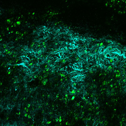
Specialized imaging windows are used to track long term (weeks to months) changes in GFP positive cancer cells (green) and second harmonic generation from fibrillar collagen fibers (cyan) in live mice after orthotopic implantation of cancer cells.
Credit: Ayrianne Crawford, Dr. Michael Hollingsworth
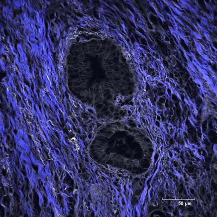
Second harmonic generation (SHG, blue) microscopy of fibrillary collagen reorganization in the tumor microenvironment of pancreatic ductal adenocarcinoma (PDAC).
Credit: Dr. Heather Jensen Smith, Dr. Tony Hollingworth, UNMC.
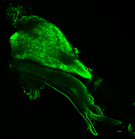
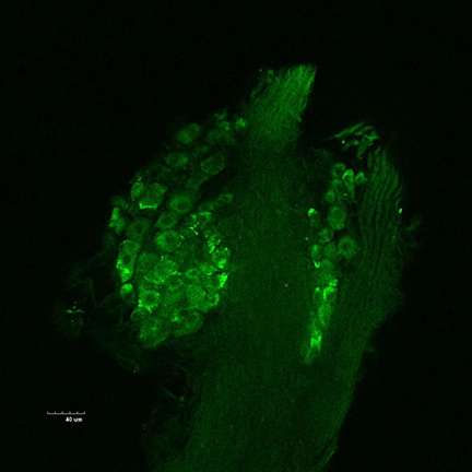
Left: Digitally stitched image of a tiled image sequence to reconstruct an optically cleared thoracic dorsal root ganglion (DRG) containing mito-roGFP mitochondria (green). 40 mm scale bar.
Right: Sequence of images obtained by optically sectioning (2 mm interval) through an optically cleared thoracic DRG containing mito-roGFP mitochondria (green).
Credit: Dr. Sharon Morais, Dr. Irving Zucker
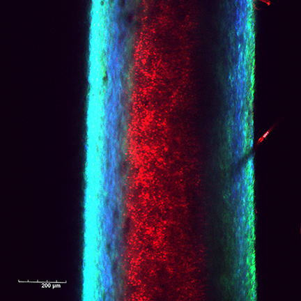
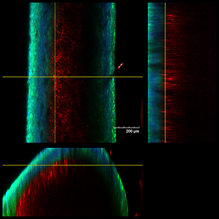
Left: Visualization of dTomato positive neutrophils (red) and collagen second harmonic (SHG) in Catchup mouse tibia.
Right: Orthogonal reconstruction of dTomato positive neutrophils (red) and collagen SHG (blue/green) in Catchup mouse tibia.
Below: Multiphoton imaging of dTomato positive neutrophil (red) movement in exposed, intact murine tibia. Collagen SHG (blue/green).
Credit: Diane Costanzo, Dr. Leah Cook
- Core Facility Directory
- Clinical Research Resources
- Nebraska Biobank
- Grant Editing Support
- Grant Resource Library
- Centers and Major Programs
- Sponsored Programs Administration (SPA)
- Comparative Medicine
- Government Relations
- Intellectual Property Assistance
- Translational Cores
- Advanced Microscopy Core Facility
- Animal Behavior Core
- Biosafety Level 3 (BSL-3) Core Facility
- Computational Chemistry
- Electron Microscopy
- Epigenomics Core Facility (ECF)
- Flow Cytometry Research Facility
- Genomics Core Facility
- Human Magnetic Resonance Imaging
- Mouse Genome Engineering Core
- Multiomics Mass Spectrometry
- Nanoimaging Core Facility
- READi Core (health informatics)
- Small Animal Imaging
- In Vivo Imaging Core Facility
- Multiphoton Intravital & Tissue Imaging (MITI) Research Core
- MRI/MRS and SPECT/CT Core Facility
- PET Core Facility
- VEVO Ultrasonic Imaging
- Translational Mouse Model Core Facility
- Core Facility Scheduling and Billing
- Backup Freezer Program