In 16-week HIV infected humanized mice, altered MEMRI signal enhancement was observed in affected brain regions including hippocampus, amygdala, thalamus, globus pallidus, caudoputamen, substantia nigra and cerebellum. MEMRI signal was coordinated with levels of HIV-1 infection, neuroinflammation (astro- and micro- gliosis), and neuronal injury. MEMRI accurately demonstrates the complexities of HIV-1 associated neuropathology in rodents that reflects, in measure, the clinical manifestations of neuroAIDS as it is seen in a human host.
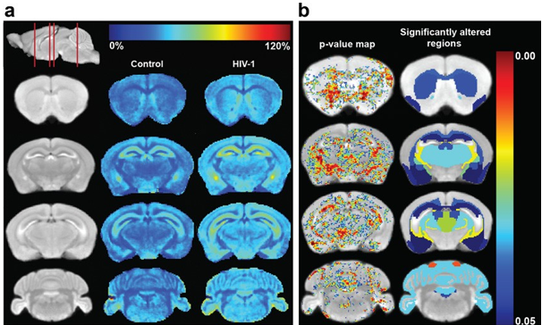
Comparison of MEMRI enhancement between HIV-1 infected animals and controls. (a) MEMRI enhancement maps. The first column (from left) shows coronal slices of the averaged MEMRI of control mice as an anatomical reference. The sagittal slice (upper left) shows respective coronal positions (red lines). The second column shows the average enhancement in control mice on the coronal slices. The third column represents the average enhancement of HIV-1 infected mice. The color bar for the enhancement maps is at the top of the figure. Dark blue color (0%) means no change in enhancement from Mn2+ compared to pre-injection signal intensity. Dark red color represents 120% signal increase compared to pre-injection. Increase in MEMRI enhancement can be seen throughout the brain of infected animals than controls. (b) Statistical comparison of MEMRI enhancement between control and HIV-1 infected animals. The left column shows the pixels with significant enhancement difference (p < 0.05) overlaid onto the averaged brain image. The color bar of p values is at the right. Dark blue color represents p = 0.05 and dark red color represents the value of 0.00. The right column shows significantly altered brain regions of infected mice using the same color scale.
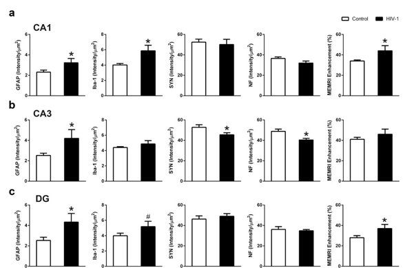
Association of immunohistology with MEMRI. (a) Quantitative analysis showed significant increase in GFAP, Iba-1, and MEMRI enhancement on CA1 region of HIV-1 infected animals compared to controls. (b) CA3 region showed significantly increased GFAP, significantly decreased SYN as well as NF, and no change in MEMRI signal in infected animals compared to controls. (c) DG region showed significantly increased GFAP, a trend of increased Iba-1, and significantly increased MEMRI enhancement in infected animals compared to controls. Data are expressed as mean SEM. (*: p < 0.05, #: p < 0.1)
For further information, see:
Aditya N Bade, Santhi Gorantla, Prasanta K Dash, Edward Makarov, Balasrinivasa R Sajja, Larisa Y Poluektova, Jiangtao Luo, Howard E Gendelman, Michael D Boska, and Yutong Liu. Manganese-Enhanced Magnetic Resonance Imaging Reflects Brain Pathology During Progressive HIV-1 Infection of Humanized Mice. Mol Neurobiol. 2016 Jul; 53(5): 3286–3297.