A 24-year-old female presents with a painful lesion.
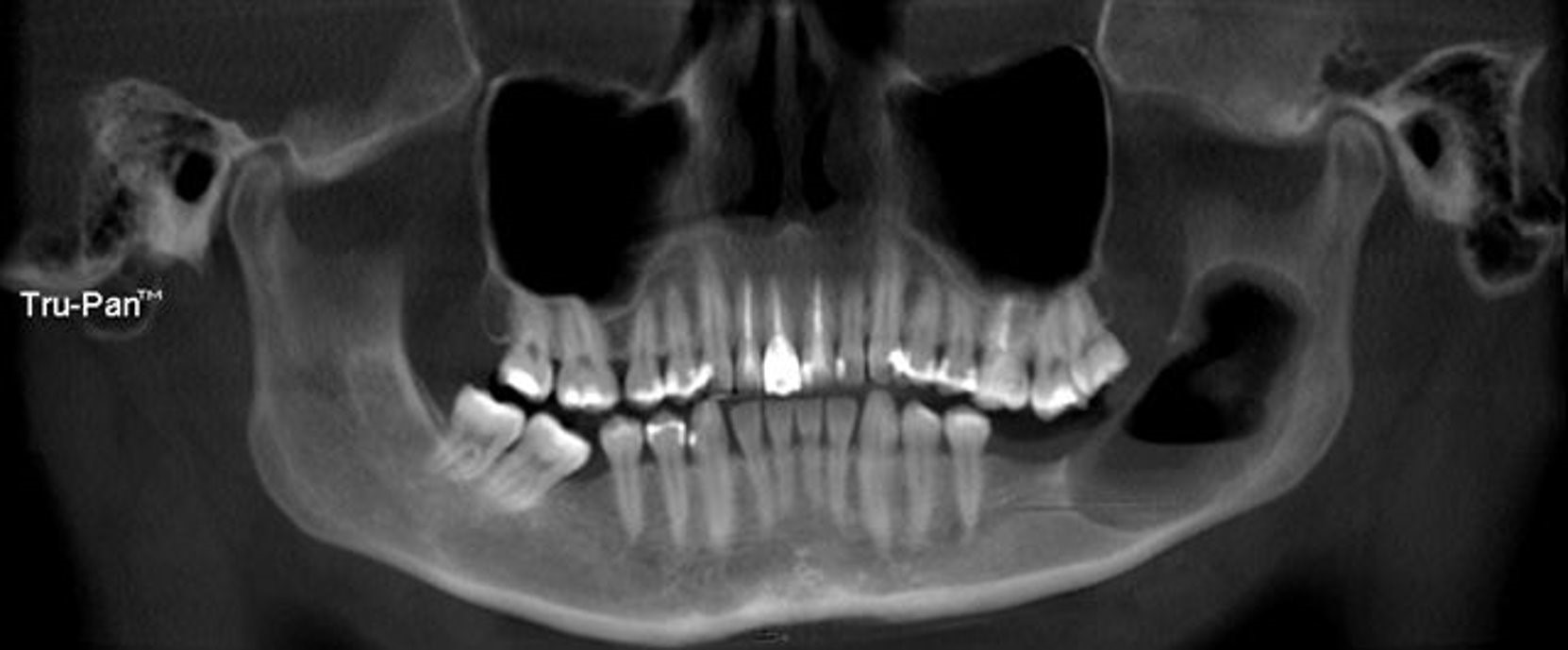
Radiographic examination: 6.0 cm x 4.0 cm radiolucency of the left posterior mandible.
History of the lesion: Pain for the past 10 months and intraoral drainage for the last 2 weeks. A mandibular tooth in the area was extracted in Guatemala 5 years ago.
Which of the following should not be included on the differential diagnosis?
Based on the histopathology, which of the following is the correct diagnosis?
What is the most appropriate management?
With what syndrome has the lesion as diagnosed in question #2 been associated?


Recent Comments