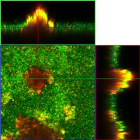
S. aureus (UAMS 1) confocal images visualized with syto 9 (live) and toto 3 (dead) stains (provided by Bayles 2010).
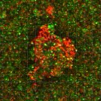
Developing S. aureus (UAMS 1) biofilm in a flow cell over a three day period (above) and video (provided by Bayles 2010).
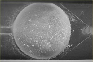
S. epidermidis mature flow cell biofilm (provided by Fey 2010).