Research Revealed: The Art of Discovery
Explore research through the eyes of the UNMC community. Research Revealed: The Art of Discovery showcases original research-related images in three categories: Hidden Worlds; Data Revealed; and Research in Action.
First, browse the entries below.
Then, vote for your overall favorite for the People's Choice Award. Voting closes Friday, Oct. 16.
A panel of judges will also select winners in the three image categories, along with Best in Show and the Emerging Researcher Award.
Winners will be announced during Research Month.
Hidden Worlds
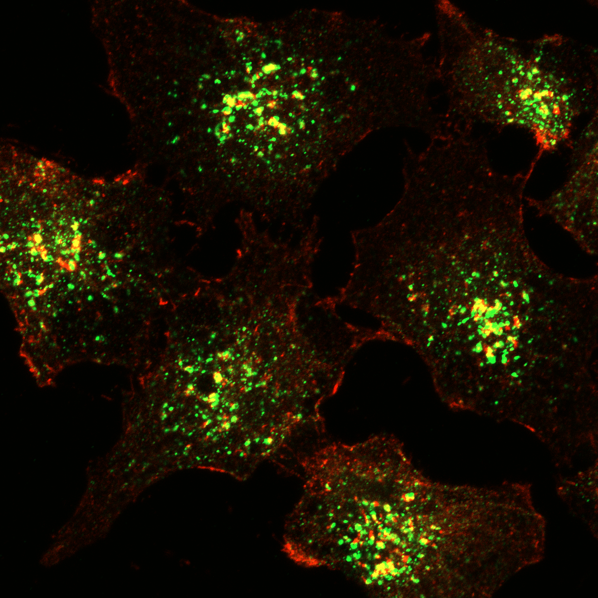
ENTRY 1: Actin association with early endosomes
Description: This image describes the impact of MICAL2-depletion on endosomes (green) and endosomal branched actin (red).
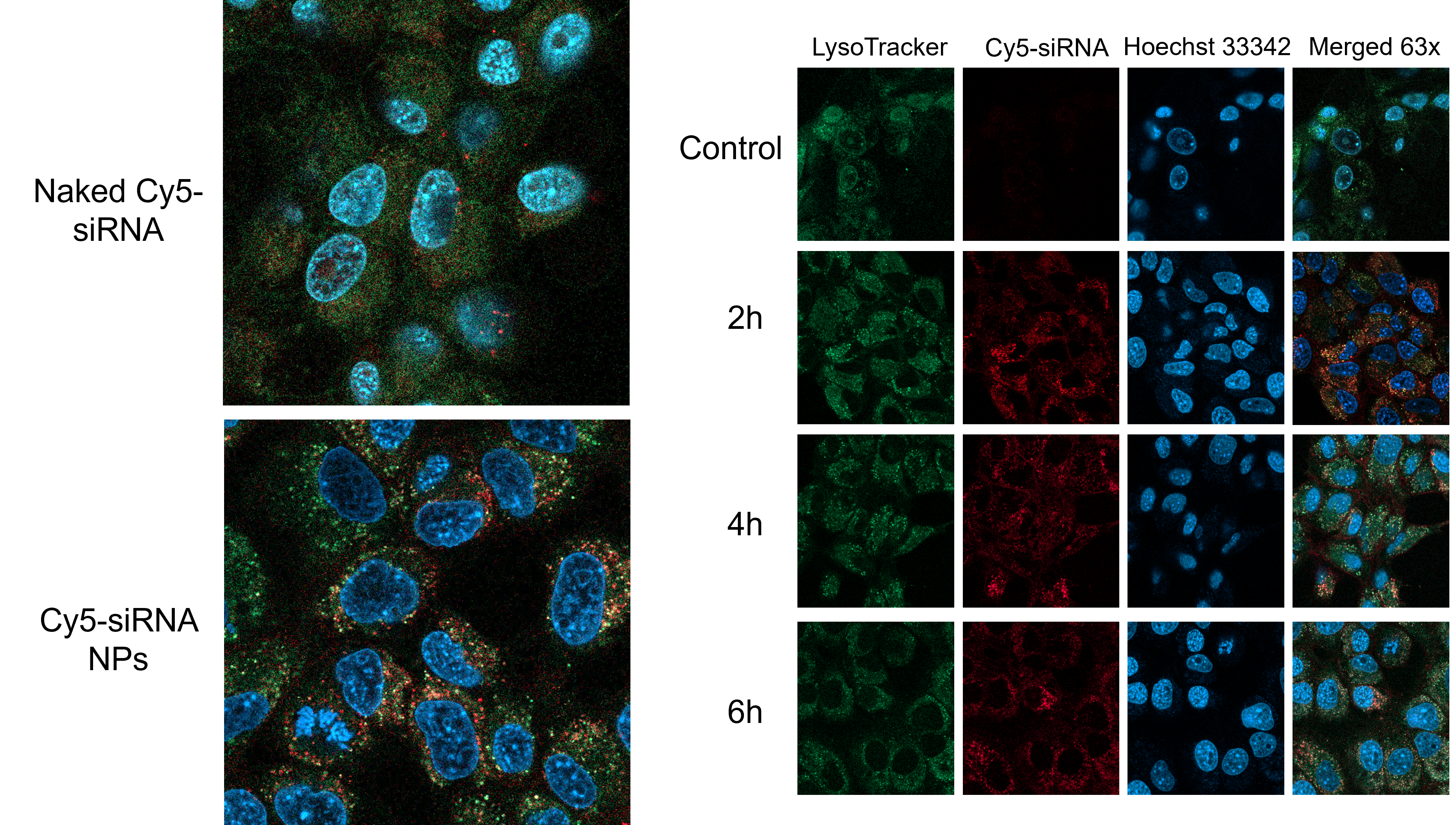
ENTRY 2: Endosomal Escape of Cy5 siRNA
Description: The image is divided into two sections. The left section shows the effect of polymeric nanoparticles on intracellular trafficking of cy5 RNA in cells; the right panel is a time-course study showing reduced co-localization of cy5 RNA from endosomes.
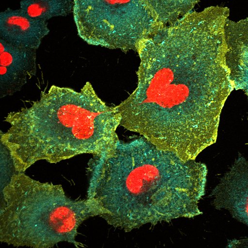
ENTRY 3: The Heart of Cellular Traffic
Description: A glimpse into lung adenocarcinoma cells, where endosomes and cell-surface receptors navigate a dynamic intracellular landscape around unexpectedly heart-shaped nuclei.
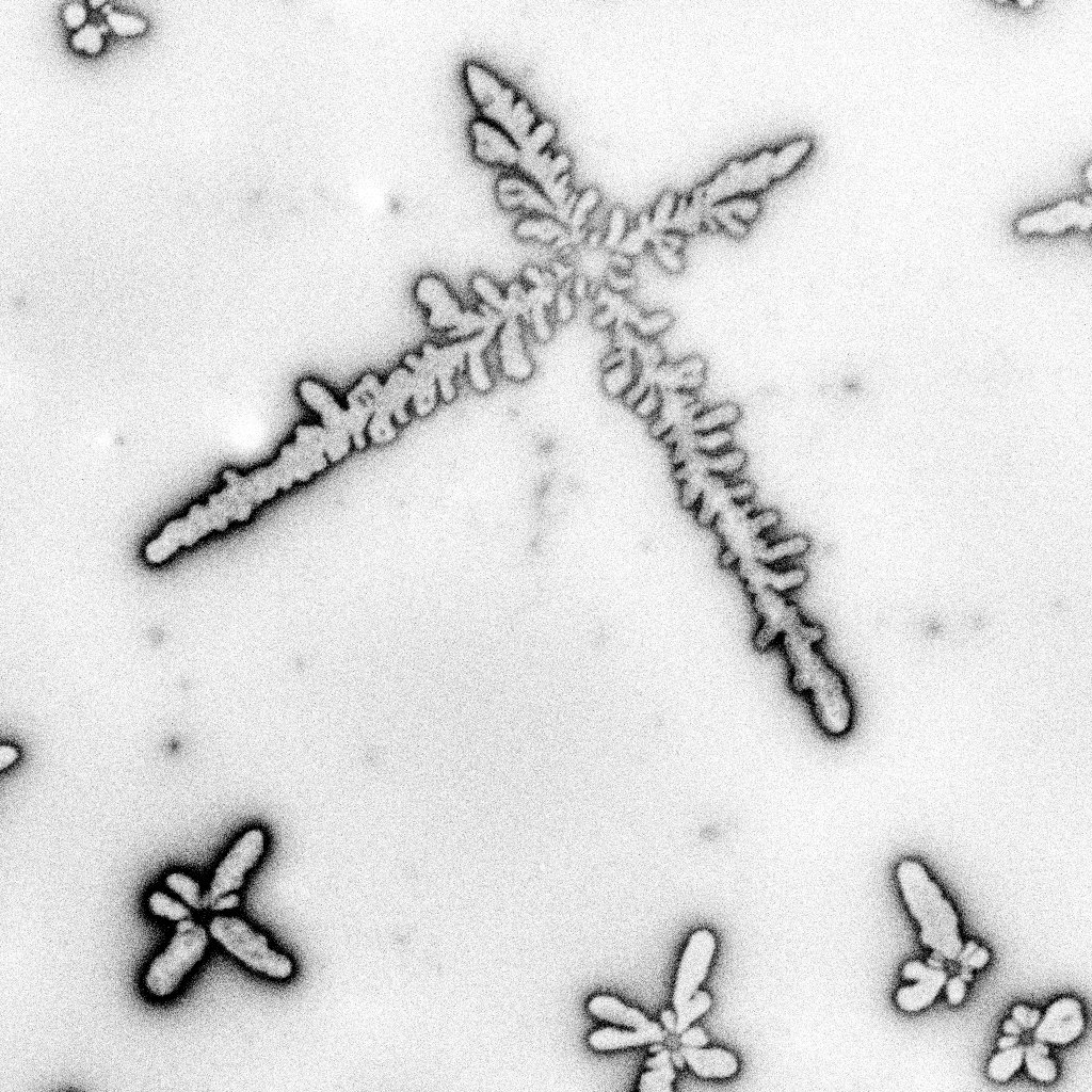
ENTRY 4: Fractals at the Interface
Description: This image captures intricate branching patterns formed through the interaction of oil and water on a microscopic surface. Although reminiscent of trees, feathers, or biological structures, these forms arise from physical processes at an interface. The image illustrates how interactions among fluids, surfaces, and molecular forces can spontaneously generate complex and highly organized patterns from relatively simple components.
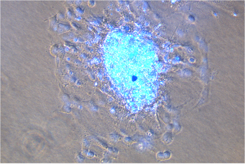
ENTRY 5: At the Edge of Invasion: A Trophoblast Spheroid in Motion
Description: This image captures a three-dimensional spheroid of human trophoblast cells as they invade outward into the surrounding extracellular matrix. The dense central spheroid serves as a starting point from which cells extend into their environment, producing the striking radial pattern visible around the core. This 3D model allows us to visualize and measure trophoblast invasion—a fundamental process by which placental cells establish the maternal–fetal interface during early pregnancy.
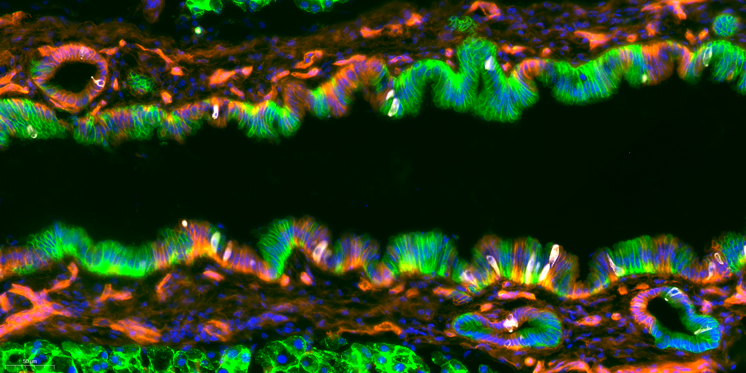
ENTRY 6: Oncogenic transformation of murine pancreas duct
Description: Main duct of a genetically engineered mouse model of PDAC utilizing a tamoxifen inducible Cre-recombinase and Cre-dependent reporter Rosa-mTmG and oncogenes. GFP+ cells represent progeny of lineage traced progenitor cells of the ductal glands with oncogenic activation. RFP+ cells represent non-lineage tagged cells. White is immunofluorescence for DCLK1 protein, marking tuft cells.
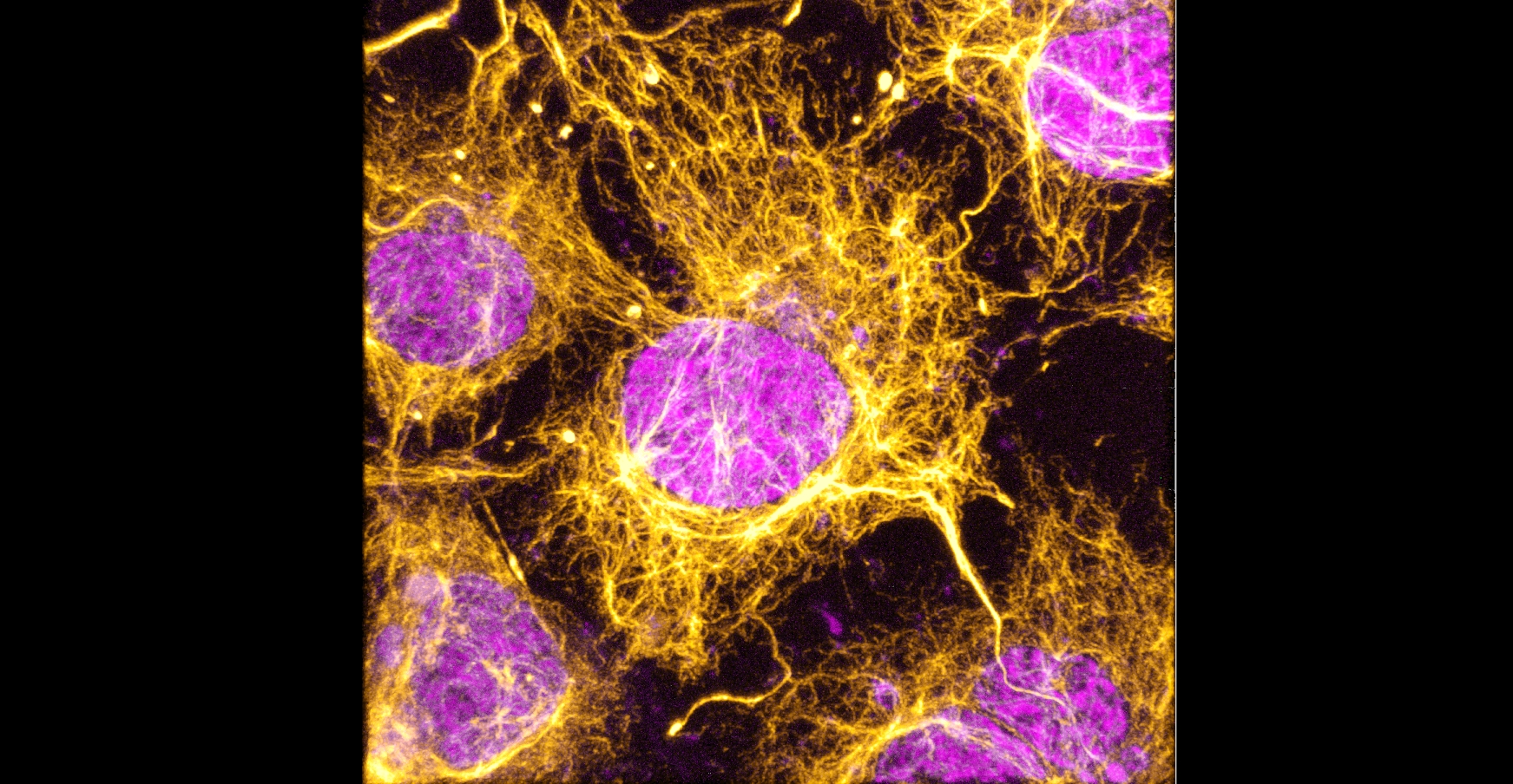
ENTRY 7: Caged in Gold
Description: This live-cell image captures the intricate vimentin network surrounding the nucleus of a live COS7 cell and extend till cell periphery. By labeling HaloTag-vimentin with a fluorescent HaloTag ligand, the architecture of this intermediate filament can be visualized and perturbed in real time.
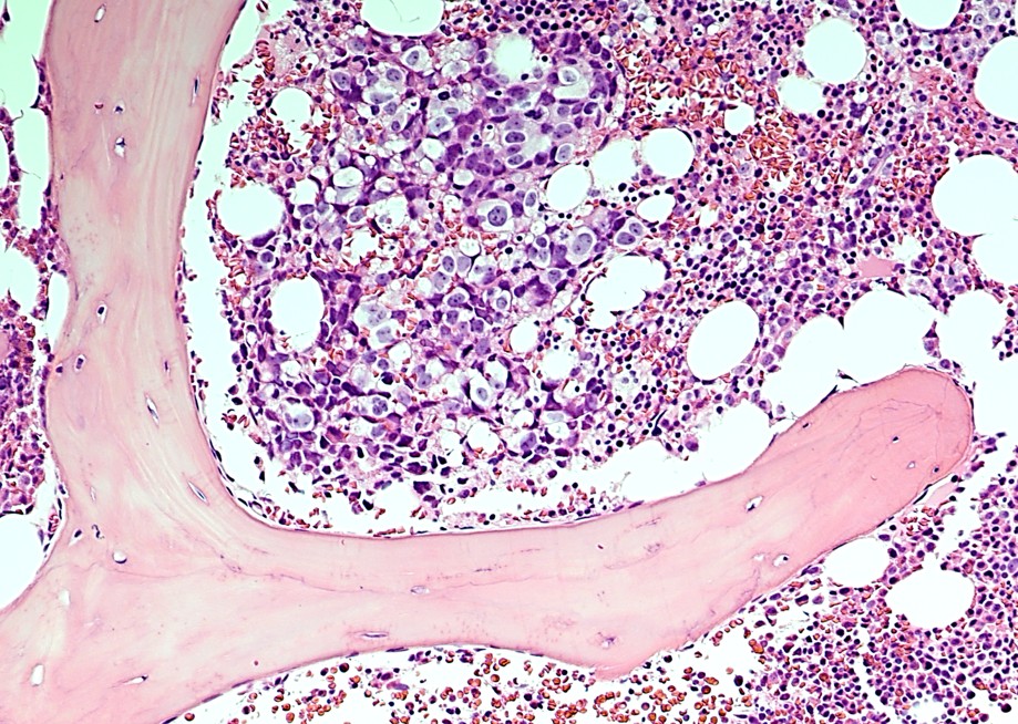
ENTRY 8: Where Tumor Meets Bone: A Microscopic Frontier
Description: This histologic image captures the microscopic interface between tumor and bone. The pink-stained trabecular bone forms a striking structural framework surrounding a densely cellular tumor region. Within the tumor, malignant cells display marked cellular and nuclear variation, creating a complex landscape of tumor cells, stromal elements, adipose spaces, and blood cells. The image provides a close-up view of the physical and cellular environment in which tumor cells interact with and potentially invade the surrounding bone.
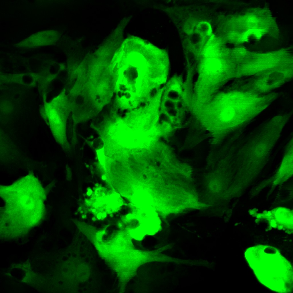
ENTRY 9: When Epicardial Cells Look Back
Description: This live-cell fluorescence image captures GFP-labeled epicardial cells during a study of CHD4-mediated regulation of cardiac development. While the experiment was designed to observe cellular morphology and organization, the cells appeared to have their own plans—several spontaneously arranged into a surprisingly face-like pattern. The GFP signal highlights the architecture of living epicardial cells, turning an ordinary imaging field into an unexpected cellular portrait.
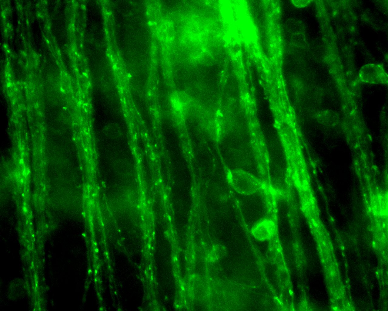
ENTRY 10: Radiant Retina
Description: Retinal ganglion cells and axons expressing Channelrhodopsin fluoresce following AAV injection into the eye of an Alzheimer mouse model. Image acquired with ThorLabs CS135MUN microscopy camera at 40X.
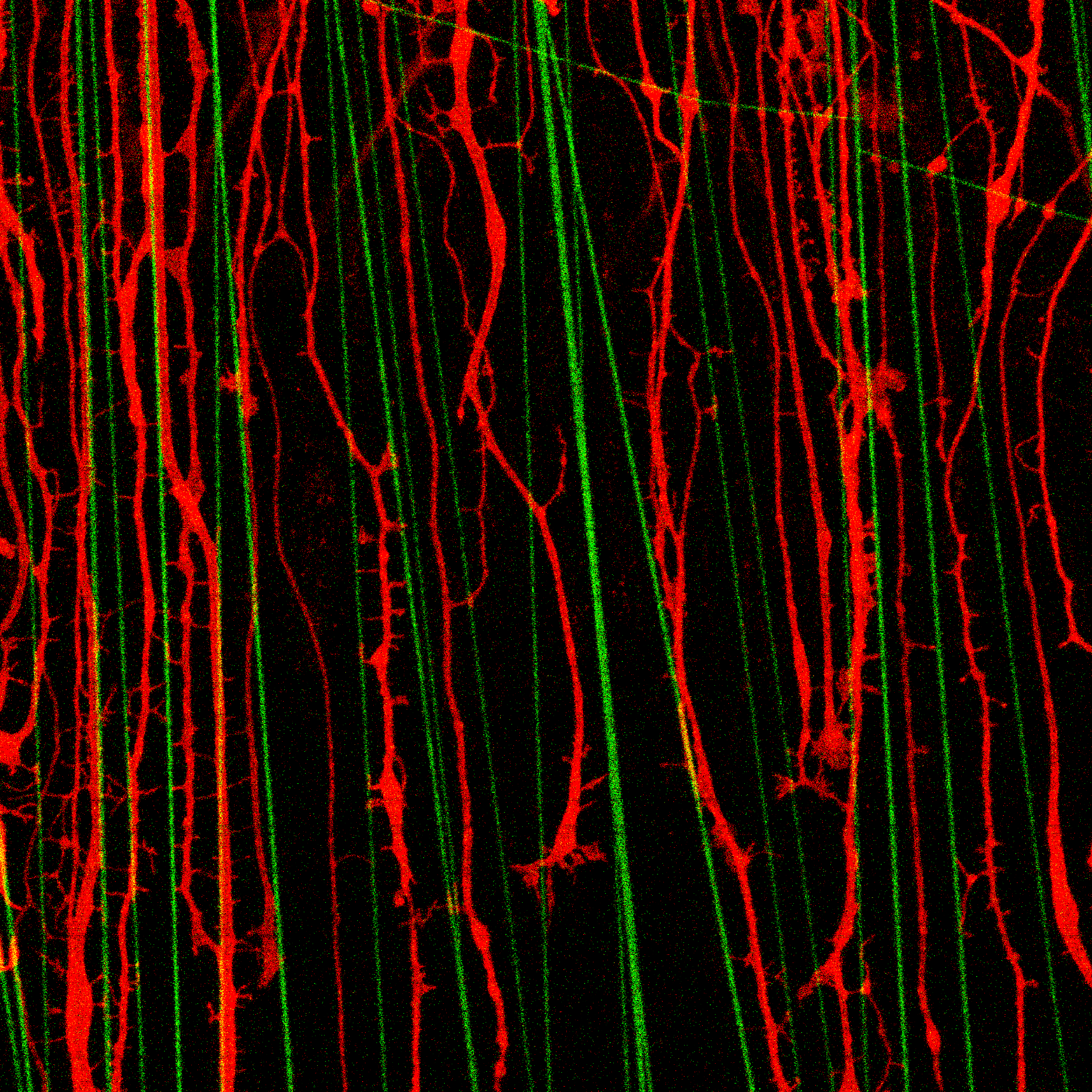
ENTRY 11: Finding the Way Back
Description: This fluorescence microscopy image captures regenerating sensory neurites (red) growing along aligned nanofibers (green) within a nerve-on-a-chip platform designed to study nerve repair. The image reveals how engineered physical guidance cues can direct neural regrowth and offers insight into biomaterial-based strategies for peripheral nerve regeneration.
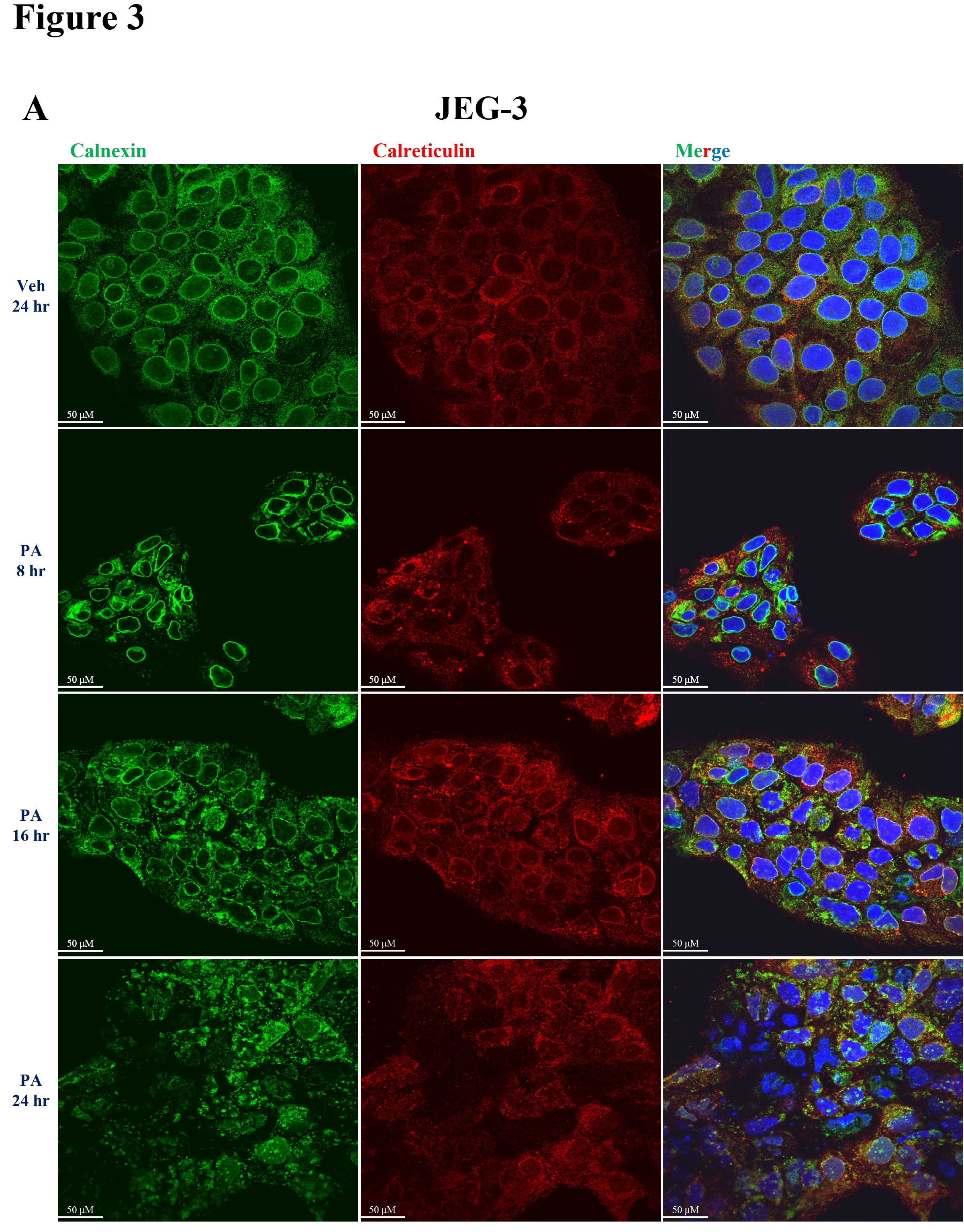
ENTRY 12: Trophoblast ER Morphological changes with Palmitate exposure
Description: JEG-3 cells treated with palmitate (PA) at 400 µM for 8-24 h showed altered ER morphology as seen with Calnexin staining compared to vehicle treated trophoblasts.
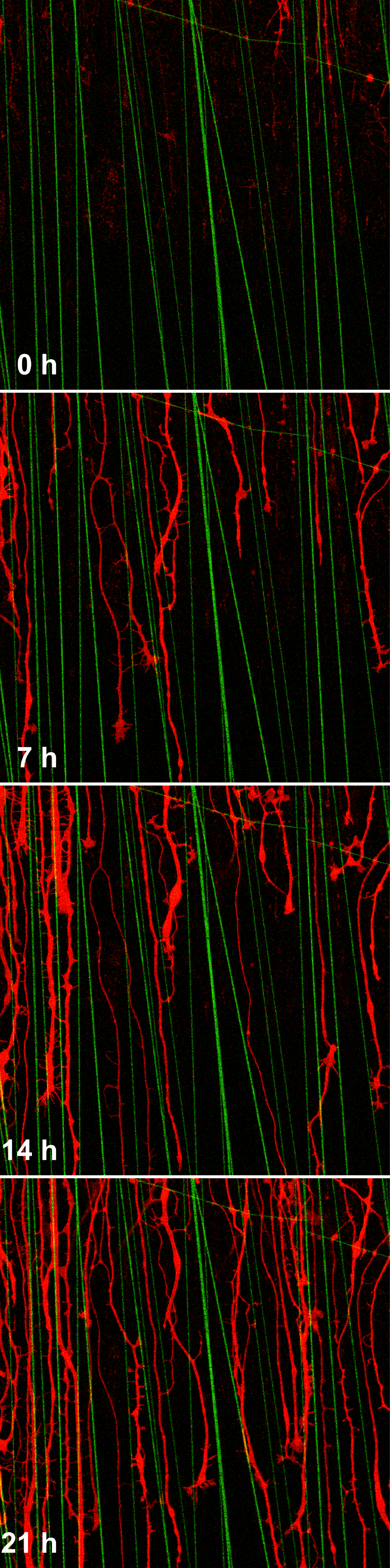
ENTRY 13: Finding the Way Back: Regeneration Over Time
Description: This time-lapse fluorescence microscopy image captures sensory neurites (red) progressively regenerating along aligned nanofibers (green) over 21 hours within a nerve-on-a-chip platform. The sequence reveals how engineered physical guidance cues can direct axonal regrowth over time, providing a dynamic view of nerve repair in a controlled microengineered environment.
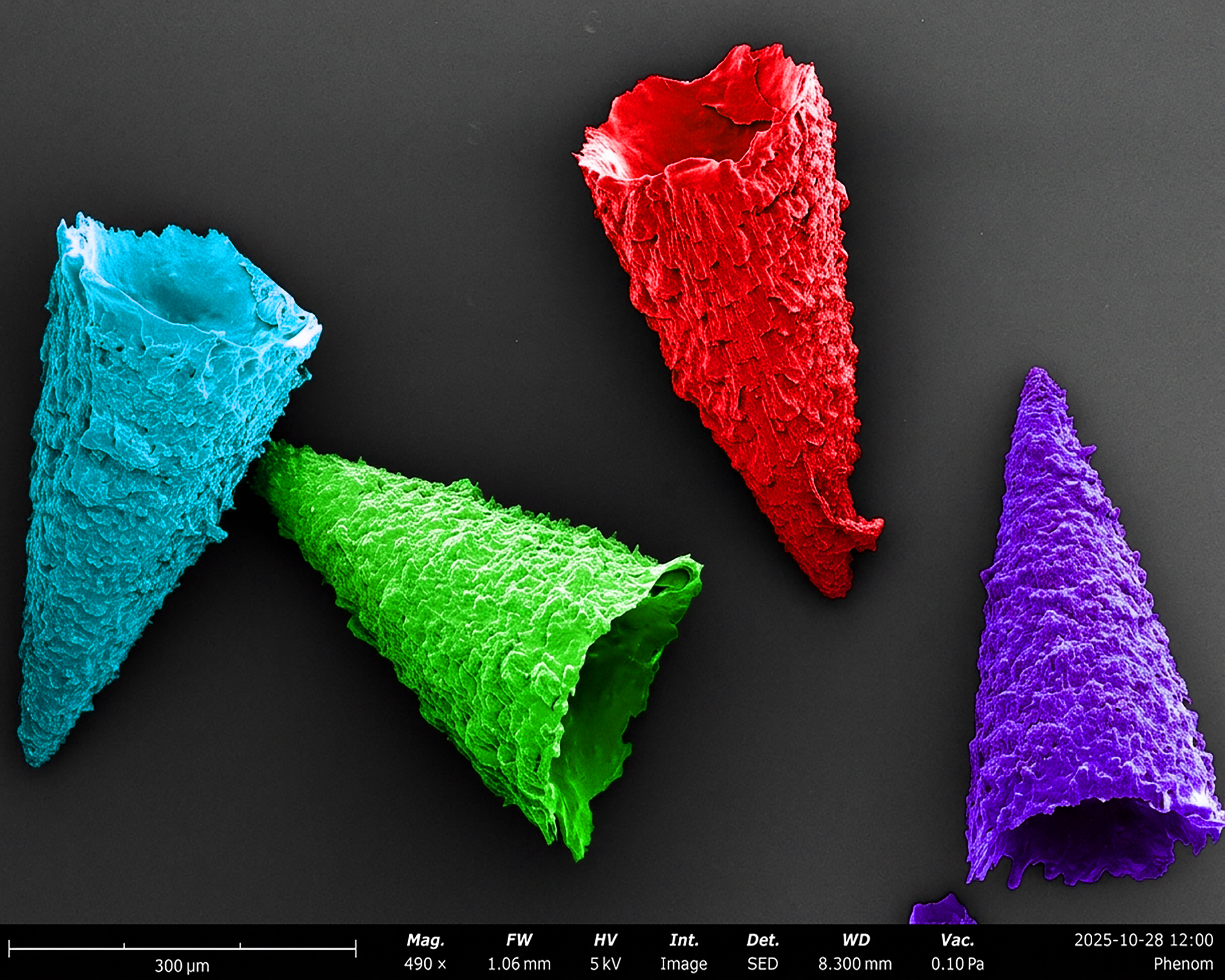
ENTRY 14: Candy-shaped Biopolymer Microneedles
Description: What appears to be colorful candies is a scanning electron microscopic (SEM) view of biopolymer microneedles, engineered for biomedical applications. This SEM image reveals individual microneedles, carefully harvested after detachment from a biofabricated microneedle patch. Each microneedle exhibits a cone-like geometry with intricate surface textures and hierarchical microstructures. The different colors are digitally applied to distinguish individual microneedles and highlight their three-dimensional architecture. Microneedles are designed to interact with the skin in a minimally invasive manner and can serve as localized platforms for transdermal and controlled delivery of therapeutic agents. (SEM Scale bar: 300 μm.)
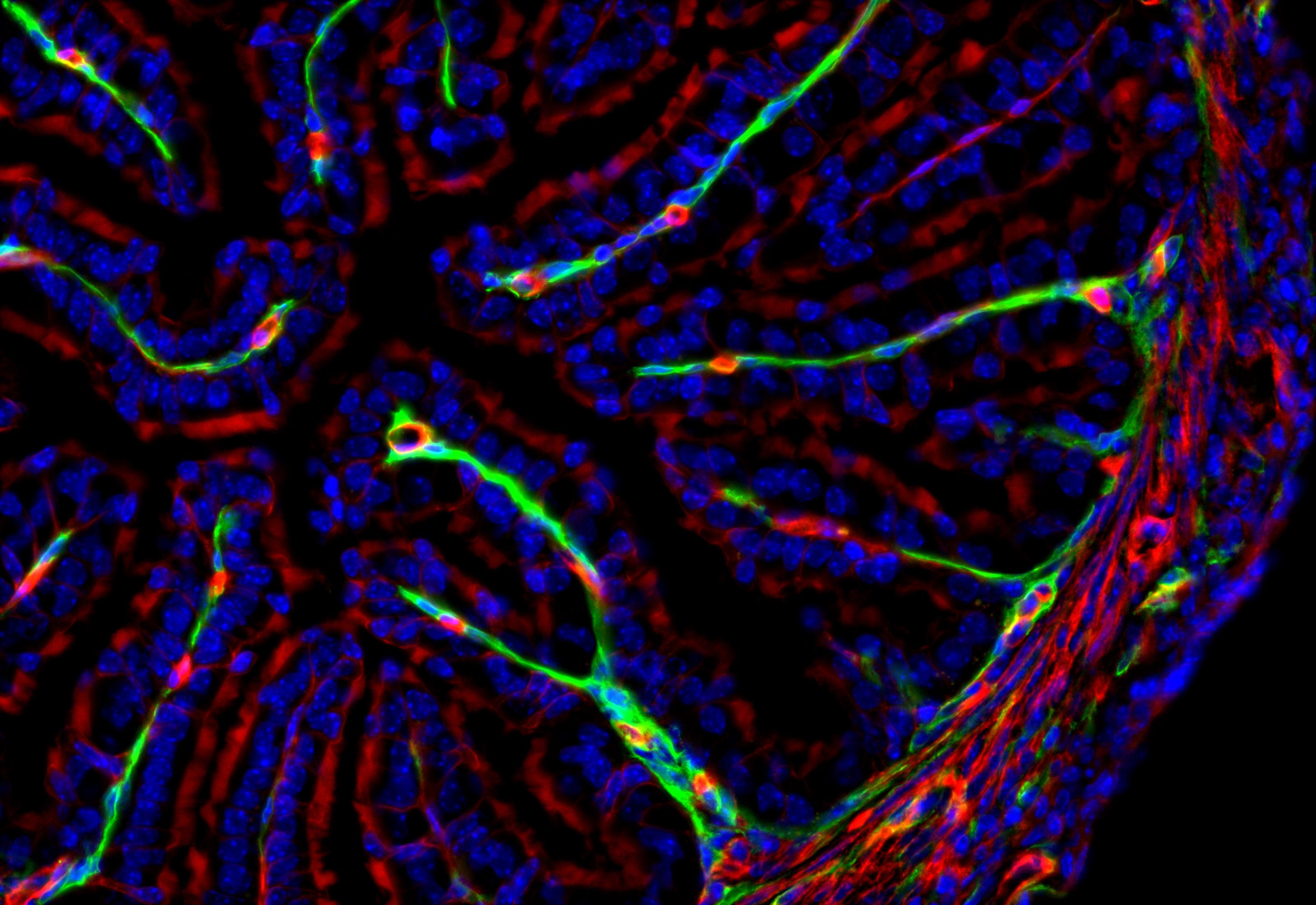
ENTRY 15: Oviduct - Relaxin Receptor Reporter
Description: Frozen section of oviduct from a dual-fluorescent relaxin reporter mouse. In this model, all cell membranes express a red fluorescent protein, except for cells expressing relaxin receptor, which express a green fluorescent protein. Green fluorescence is visible in the inner mucosa and in the muscularis layer.
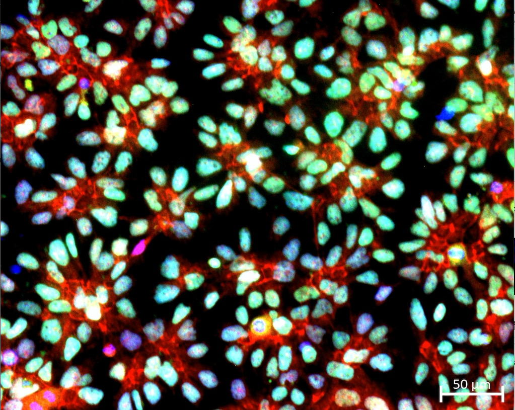
ENTRY 16: Nuclear co-expression of the transcription factors PAX6 (green) and RX (red) in retinal progenitor cells (RPCs)
Description: Double immunofluorescence staining showing nuclear co-expression of the eye-field transcription factors PAX6 (green) and RX (red) in human embryonic stem cell (hESC)-derived retinal progenitor cells (RPCs) cultured as a monolayer.
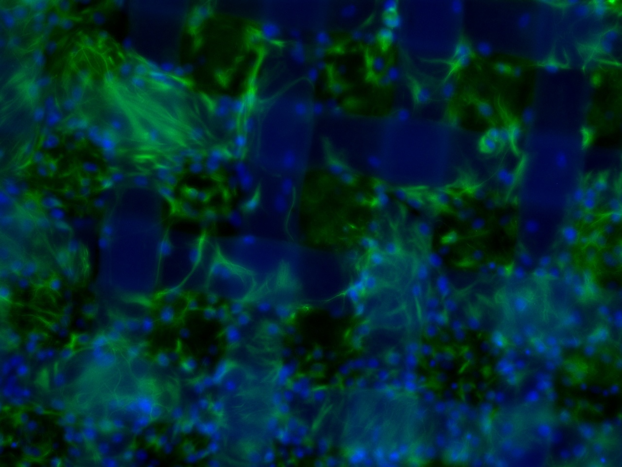
ENTRY 17: Plaid Astrocytes
Description: Mouse organotypic brain slice cultures were developed to model neonatal hypoxic-ischemic brain injury. This brain slice was cultured on a woven insert and the astrocytes thrived, growing through the mesh and securing the slice to the culture insert, as demonstrated by the cells stained with GFAP (green) and nuclei labelled with Hoechst (blue).
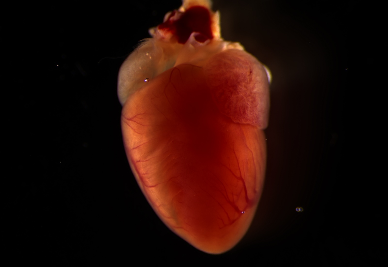
ENTRY 18: The Heart of Life
Description: This image captures an intact mouse heart, highlighting the beauty and complexity of an organ essential for sustaining life.
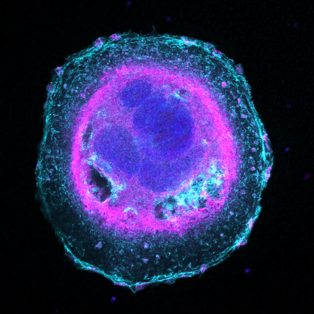
ENTRY 19: Concentric
Description: This is a single CFPAC-1 pancreatic cancer cell, imaged by confocal microscopy (Zeiss LSM 800 with Airyscan, 63x objective). Ras is shown in light blue, PIP5K1A in pink, and DNA (DAPI) in dark blue. Ras traces the cell's outer edge and fills smaller pockets near the nuclei, while PIP5K1A forms a bright perinuclear ring and appears as tiny, dense clusters in the cytoplasm and at the plasma membrane. The nuclei sit at the core of the cell. Layer by layer, the visualization of Ras, PIP5K1A, and DNA creates a planetary-like structure, with every detail having its own defined place. This cell is remarkably large and multinucleated, a feature seen in pancreatic cancer cells with genomic instability.
Data Revealed
ENTRY 1: Bacterial cell walls
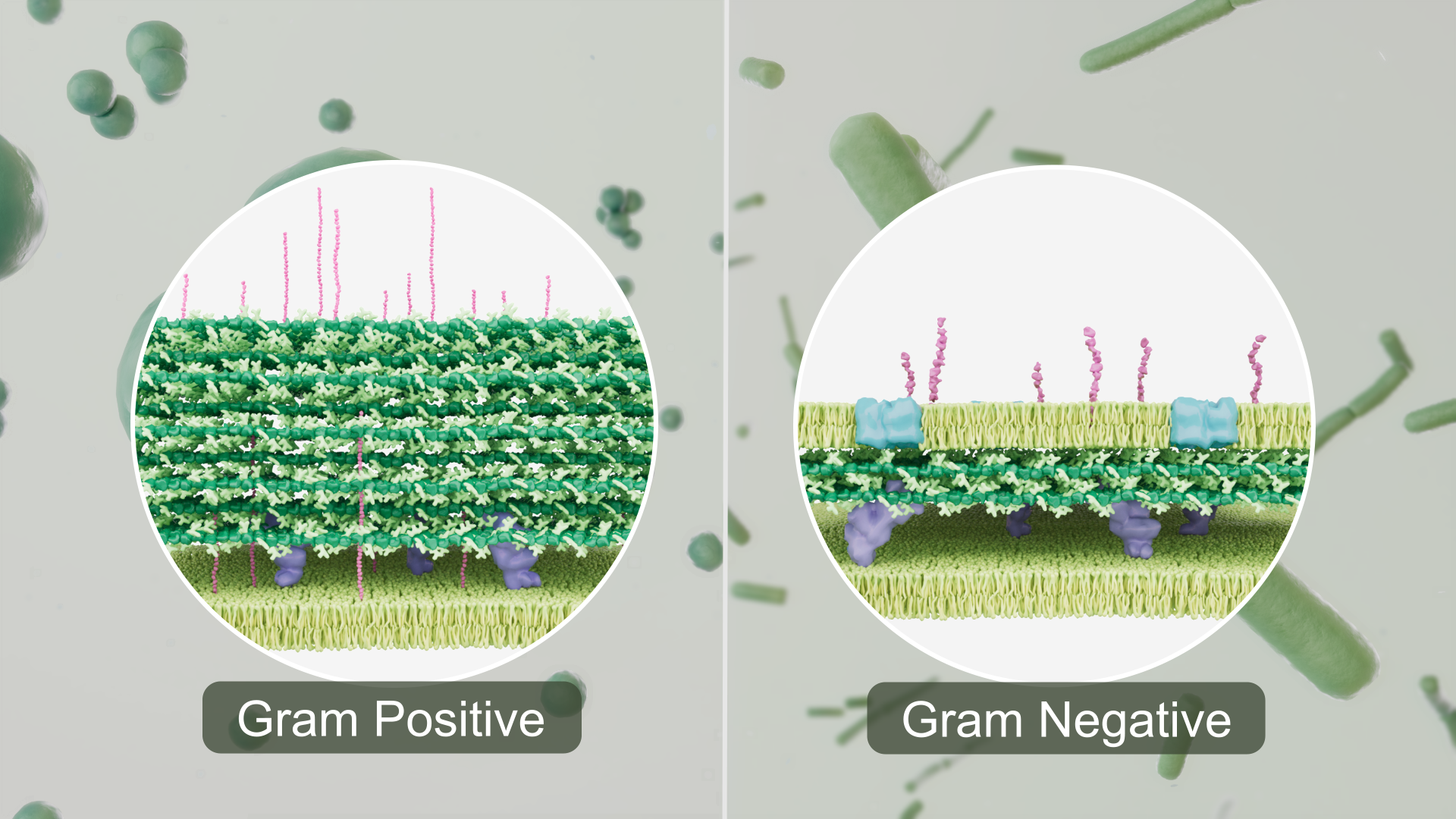
Description: This image is a still from an educational animation explaining the mechanism of Gram staining. The insets show a visual representation of the structures of cell walls in Gram-positive (left) and Gram-negative (right) bacteria.
The Gram-positive cell wall contains lipoteichoic and teichoic acids (pink), peptidoglycan (green), penicillin-binding protein (violet), and a plasma membrane (lime green).
The Gram-negative cell wall contains lipopolysaccharides (pink), an outer membrane (lime green) with porins (blue) embedded in it, peptidoglycan (green), penicillin-binding protein (violet), and a plasma/inner membrane (lime green).
The 3D molecular models were reconstructed from data from the RCSB Protein Data Bank, simulated models of bacterial peptidoglycan layers, and visual reconstruction from published literature. Visual Molecular Dynamics was used to load and visualize molecular structures, and the entire cell wall was assembled in Cinema4D. In reality, these structures would be more numerous and densely arranged. For example, the peptidoglycan layer of the Gram-positive cell wall would be considerably thicker. For educational clarity, the number of molecules and layers was reduced.
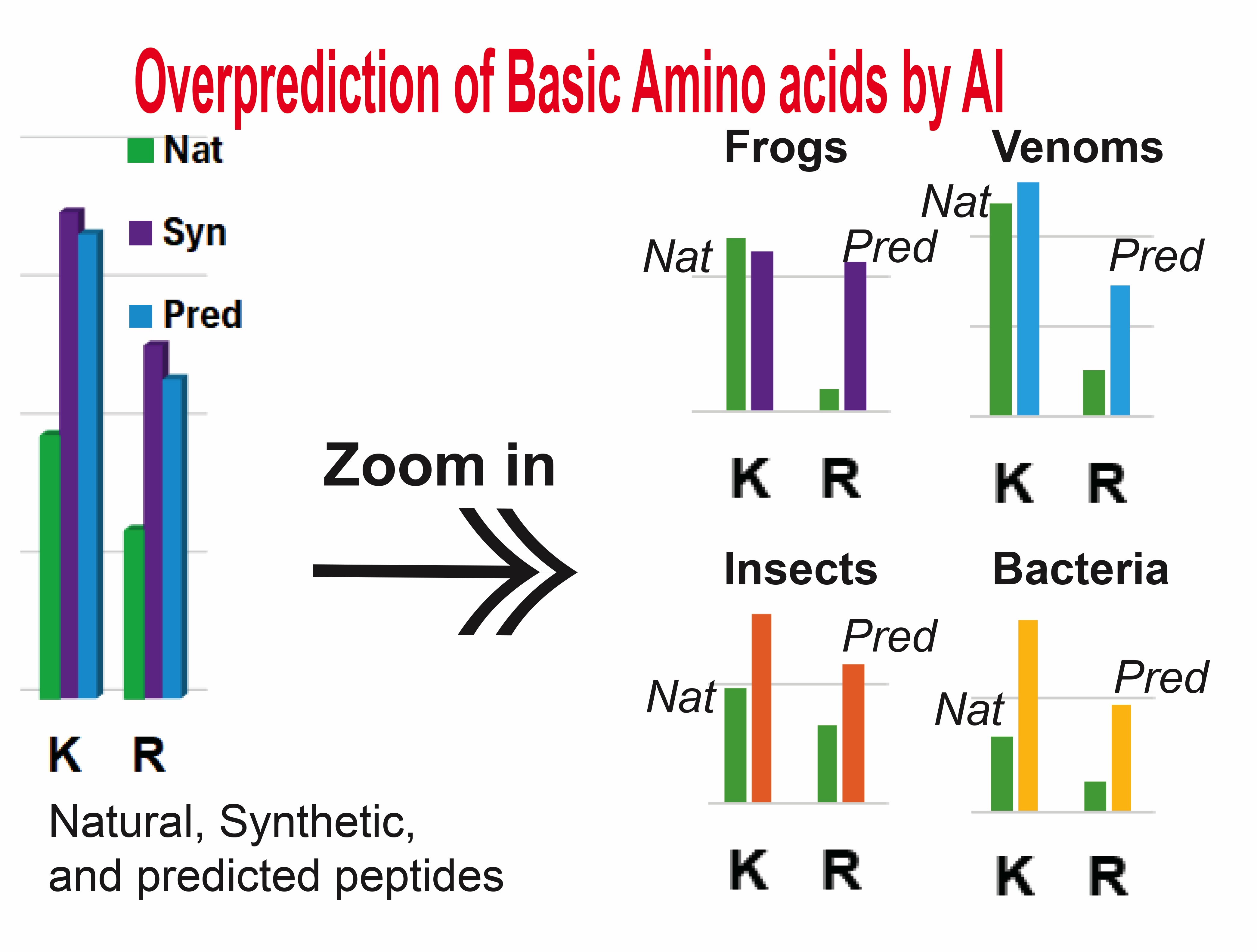
ENTRY 2: Current genomic predicted antimicrobial peptides have deviated from natural profiles and resemble man-made peptides
Description: The antimicrobial peptide database (APD; https://aps.unmc.edu) is widely used worldwide for AI prediciton of antimicrobial peptides. Our database finding displayed in the image underscores the importance of right data sets for unbiased prediction in the future. Since the inclusion of a "large" artificial sequences might be problematic, it can be anticiapted that the natural antimicrobial peptides accumulated in the APD for over 23 years will play an essential role in predicting such peptides from sequenced genomes.
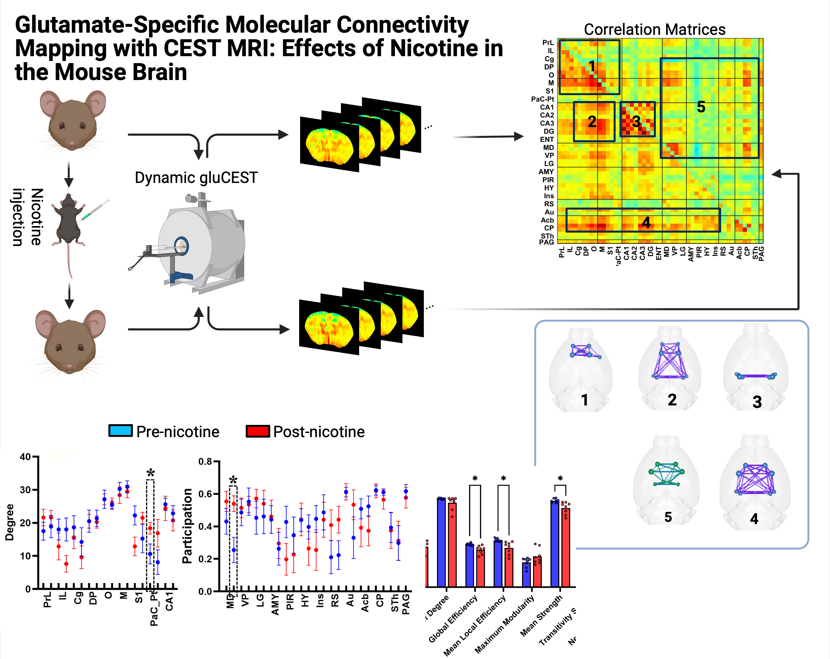
ENTRY 3: Brain Glutamate Networks and Nicotine-Induced Changes Detected by MRI
Description: This is a dynamic glutamate chemical exchange saturation transfer (gluCEST) MRI study of nicotine effects on mouse brain glutamate-related connectivity. Mice were scanned before and after nicotine injection, producing serial gluCEST maps. Regional gluCEST signals were then correlated to generate a brain-region correlation matrix and identify five connectivity modules, shown as network diagrams.
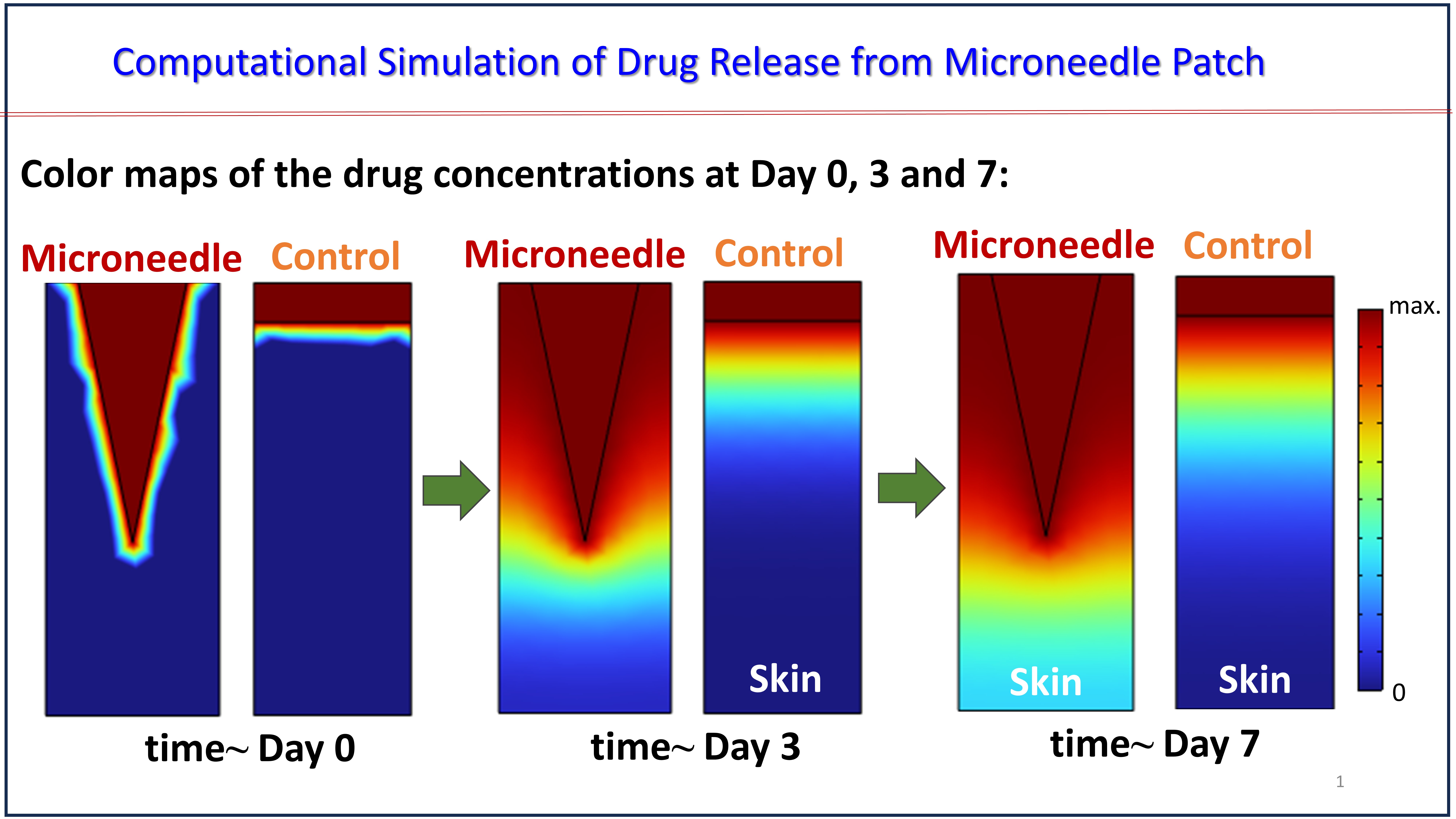
ENTRY 4: Mapping the Invisible: Computational Modeling of Microneedle Drug Diffusion
Description: This computational simulation visualizes the spatiotemporal transport of a therapeutic payload from a microneedle patch compared with a conventional topical control over 7 days. Using finite element modeling of drug diffusion and transport through skin tissue, the time-series color maps reveal evolving drug concentration gradients. At Day 0, the microneedle geometry enables localized delivery beneath the stratum corneum, while the topical control initially confines the modeled payload to the skin surface. By Days 3 and 7, the simulations illustrate the progressive, multidirectional diffusion of the therapeutic agent into deeper tissue regions. By transforming complex transport equations into intuitive visual maps, this comparison reveals how delivery geometry may influence the spatial distribution and temporal evolution of a therapeutic payload in skin.
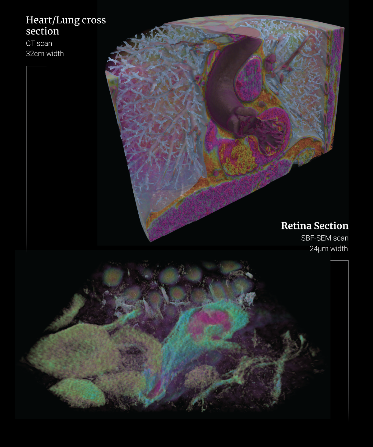
ENTRY 5: Volumetric Scans: Big and Small
Description: These images were rendered in WonderVol, an app that we are developing at iEXCEL for interactively viewing various types of volumetric scans on multiple kinds of devices. The upper image shows a human-scale region, while the lower image shows a microscopic subject. Both have been colorized and rendered in 3D using the same tool.
Research in Action
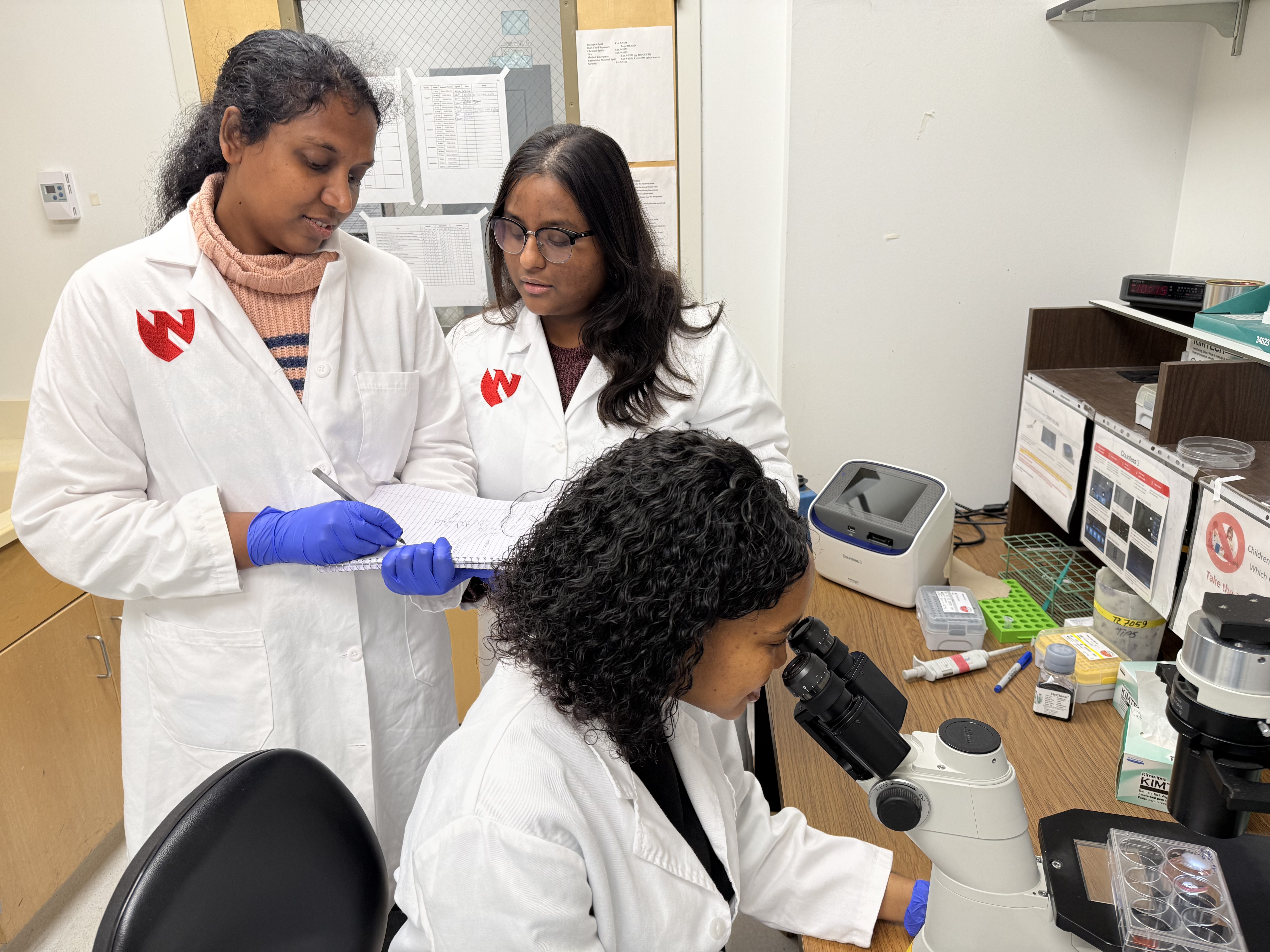
ENTRY 1: Cancer Cell Culture, Experimental Analysis, and Microscopic Visualization
Description: Researchers discussing ongoing cancer cell culture experiments and examining cells under a microscope.
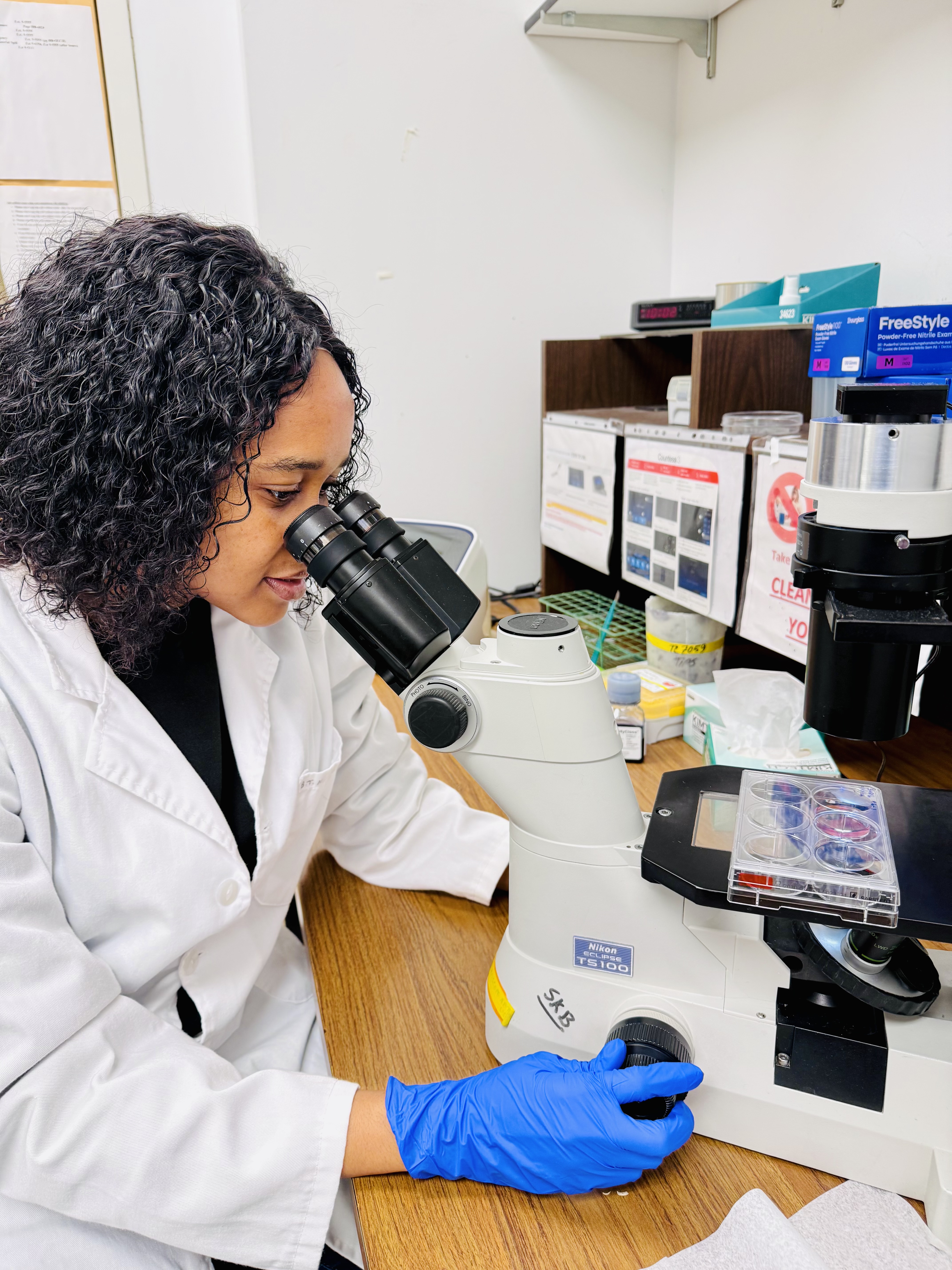
ENTRY 2: Decoding Cancer, One Cell at a Time
Description: This image captures a researcher examining pancreatic cancer cells under a microscope, highlighting the hands-on work that drives scientific discovery.
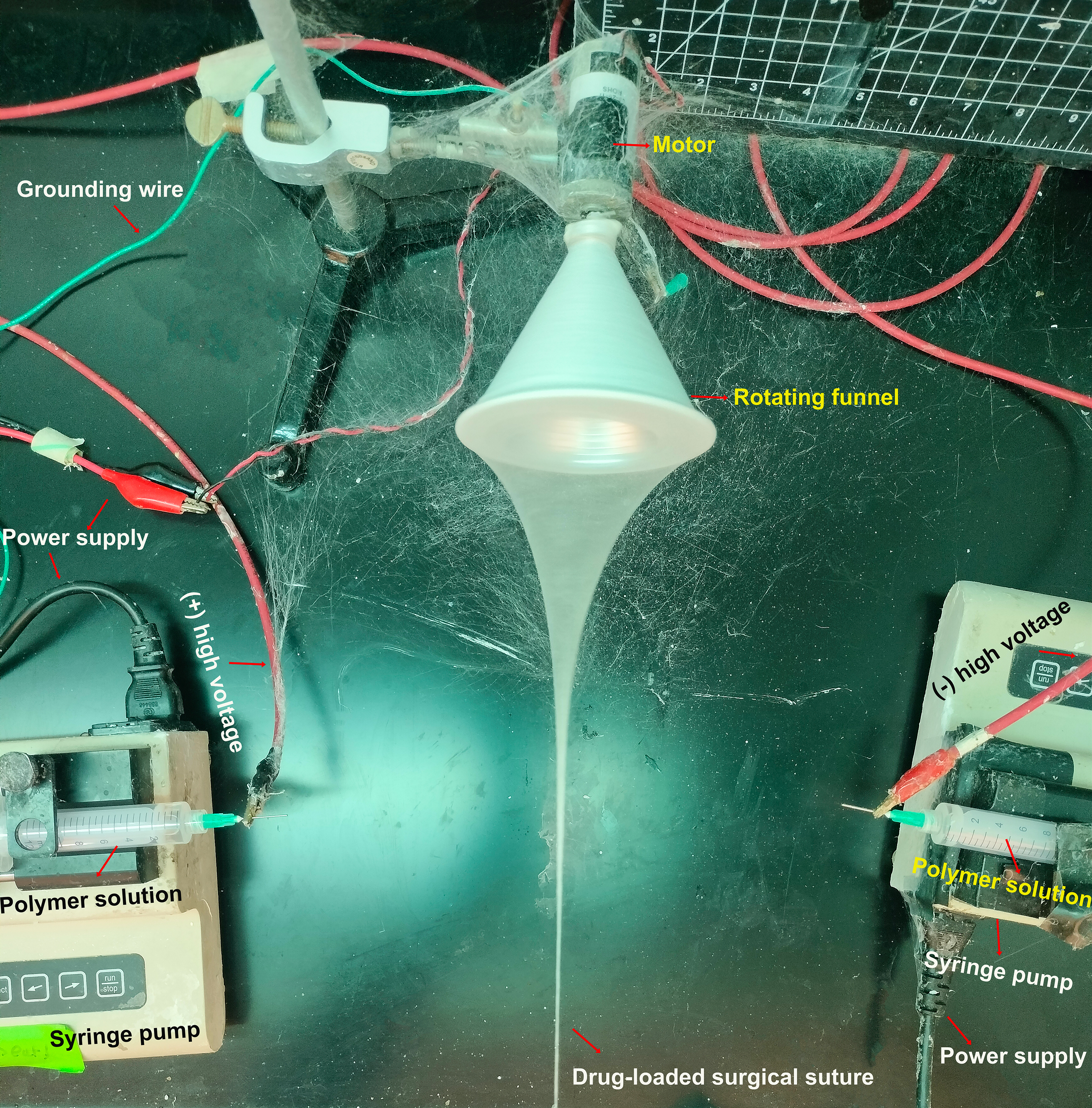
ENTRY 4: The Art of Healing: Spinning the Future of Surgical Sutures
Description: This photograph captures a custom-built experimental setup designed to explore the fabrication of drug-delivering surgical sutures using electrohydrodynamic fiber processing. A motor-driven rotating funnel and opposing high-voltage electrodes are integrated into this electrospinning system to investigate polymer nanofiber formation and the development of a surgical suture structure. The setup illustrates the intersection of physics, nanoengineering, and biomaterials research. The image was captured during laboratory experimentation and documents the apparatus and fiber-forming process.