
Advanced Microscopy
Some questions can only be answered by looking. Microscopy shows where molecules, cells and structures actually sit—from detail finer than a wavelength of light, up to whole intact organs rendered in three dimensions.
The Advanced Microscopy Shared Resource gives researchers access to a full range of imaging systems, from super-resolution and confocal microscopes to whole-slide scanners and light sheet microscopes for intact tissue. Imaging specialists can help plan experiments, prepare samples, collect images and analyze the results.
New Project Consultation
Starting a new project involving one or more Shared Resources, or interested in learning more about available services? Contact Dr. Kate Hyde to schedule a consultation or complete the consultation form in Stratocore PPMS.
Request Service
Use Stratocore PPMS to request core facility services and manage scheduling and billing.
Pricing Benefits
Subsidized pricing is available to Fred & Pamela Buffett Cancer Center members. If you haven't received an email about subsidized pricing, please get in touch with cancer center administration to see if you qualify.Contact: buffettcancercenter@unmc.edu
If you conduct cancer-related research at UNMC and are not yet a member of a Cancer Center research program, visit the Fred & Pamela Buffett Cancer Center Membership page to learn more about becoming a member.
Support at Every Stage
Consultation. Talk with imaging specialists before you start. Choosing the right instrument, preparing the sample, and picking labels are the decisions that most determine whether your experiment works.
Training and continued learning. Specialists train researchers to run most systems independently. The core maintains sample preparation guides, protocols, and recorded workshops for users at any experience level, runs regular workshops with instrument vendors, and keeps users informed as instruments and software are updated.
Sample preparation. The core can assist with preparations such as tissue clearing, a required step before most whole-tissue 3D imaging.
Imaging. Staff can collect your images for you, work alongside you, or set you up to run the instrument yourself. Access varies by instrument and is noted below.
Image analysis. Dedicated workstations with specialized software are available by request.
Data storage. Images are transferred to the core’s managed server, which helps meet NIH data management and sharing requirements.
Advanced Microscopy Shared Resource Instruments
The Advanced Microscopy Shared Resource provides access to a range of advanced imaging instruments that support cancer research across the Fred & Pamela Buffett Cancer Center. The instruments offer researchers tools for high-resolution imaging, visualization and analysis of biological samples. Explore the instruments below to learn more about their capabilities and applications. Instruments are grouped by the scale of what they can resolve, from the very small to whole intact tissues.
| Imaging Type | Description | Instrument Available | Additional Instruments Available |
|---|---|---|---|
| Super-Resolution Imaging | Super-resolution microscopes see detail finer than a standard light microscope can physically resolve, revealing structures inside cells that would otherwise blur together. These methods trade speed for detail: many images are collected and combined, so they suit fixed samples and bright, stable labels. | Zeiss Elyra PS.1: Supports several super-resolution methods (SIM, PALM, STORM, TIRF), reaching roughly 120 nm resolution with structured illumination and as fine as ~20 nm with single-molecule methods. Images up to four colors. Staff-operated; experienced users may qualify for independent access. | N/A |
| Confocal Imaging | Confocal microscopes use a pinhole to reject out-of-focus light, giving clean optical “slices” through a sample that can be stacked into a 3D image. These are the everyday workhorses for co-localization, live-cell, 3D, and time-lapse imaging. | Zeiss LSM 800 with Airyscan: An array of detectors sharpens conventional confocal resolution from ~250 nm to ~180 nm. Sensitive detectors allow lower laser power, meaning less bleaching and less damage to live cells. Includes an incubator for live-cell work. Training available for independent use. | N/A |
| Whole Slide Imaging | Whole-slide scanners digitize entire tissue sections, so you can quantify staining across the whole specimen instead of hand-picking fields of view. | Zeiss Axioscan 7: Scans up to 100 slides in a run, in fluorescence or brightfield, supporting standard IHC and immunofluorescence as well as cyclic immunofluorescence. Staff-operated; researchers complete a sample submission form before submitting slides. | N/A |
| Whole Tissue (3D) Imaging | Light sheet microscopy illuminates a sample from the side, one thin plane at a time, so intact tissues can be imaged in three dimensions tens to hundreds of times faster than with a confocal microscope — but the tissue must first be made transparent. | Miltenyi Ultramicroscope II Light Sheet Microscope: Volumetric imaging of intact tissues, small organs, and whole small organisms (samples up to roughly 1.5 cm cubed). Resolution of ~3 µm and above, the trade-off for imaging large volumes. Training available for independent use. X-Clarity Tissue Clearing System: Makes fixed tissue optically transparent by removing the lipids that scatter light, while preserving tissue structure and fluorescent labels. Staff-operated. | X-Clarity Tissue Clearing System: Makes fixed tissue optically transparent by removing the lipids that scatter light, while preserving tissue structure and fluorescent labels. Staff-operated. |
| Image Analysis | Convert complex microscope images into measurable scientific insights. | HALO: Cell-by-cell quantification across whole tissue sections, with modules for spatial relationships between cells, multiplexed marker analysis, FISH/RNAscope, machine-learning tissue classification, and serial section registration. Imaris: 3D and 4D reconstruction, segmentation, and tracking, particularly suited to super-resolution and light sheet datasets. Also available: Zen Black, QuPath, and CT-FIRE/CurveAlign. | Imaris: 3D and 4D reconstruction, segmentation, and tracking, particularly suited to super-resolution and light sheet datasets. Also available: Zen Black, QuPath, and CT-FIRE/CurveAlign. |
| Preclinical Imaging | Multiphoton microscopy uses long-wavelength light that penetrates deep into dense tissue with less damage, making it possible to image inside living animals. It can also detect collagen without any labeling. | Olympus FVMPE-RS Multiphoton Microscope: Built for deep-tissue and live-animal (intravital) imaging, with a tunable near-infrared laser. Fast resonant scanning captures rapid changes in living tissue, and second-harmonic generation imaging maps collagen structure label-free. Sensitive detectors and a heated stage support extended intravital sessions. Neurotar Mobile Home Cage: Allows imaging of a live, awake, moving animal while tracking behavior. | Neurotar Mobile Home Cage: Allows imaging of a live, awake, moving animal while tracking behavior. |
Citing Shared Resources
Researchers using the Fred & Pamela Buffett Cancer Center Shared Resources for publications, presentations or other scholarly outputs are required to acknowledge the use of them. Proper acknowledgment helps support the continued availability and funding of these cores.
Please acknowledge the Cancer Center Support Grant (P30 CA036727) in all publications and abstracts that have used these services.
Suggested language: Research reported in this publication was supported by the Advanced Microscopy Shared Resource (RRID:SCR_022467) and the National Cancer Institute under award number P30 CA036727. The content is solely the responsibility of the authors and does not necessarily represent the official views of the National Cancer Institute.
Director
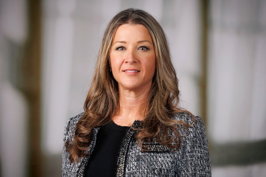
Heather Jensen-Smith, PhD
Director, Advanced Microscopy Shared Resource
Durham Research Center 1056H | Send Email | 402-559-6659