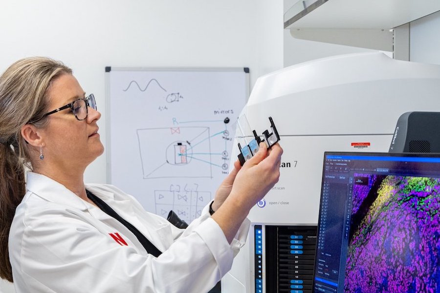
Advanced Microscopy Shared Resource
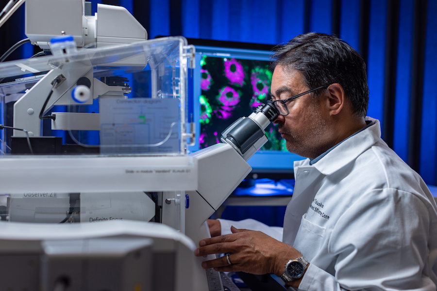
Supported by the Fred & Pamela Buffett Cancer Center (FPBCC), the Advanced Microscopy Shared Resource serves as a hub for cutting-edge technologies, expertise, and training, driving collaborative innovation across various research areas. It provides a comprehensive range of biomedical imaging modalities capable of capturing intricate biological processes at different scales, from single molecules (nanometer, super-resolution) to sub-cellular and cellular levels (micron, confocal, whole slide scanning), and even tissue or small organs (millimeter, light sheet). The facility offers instruction and user access to advanced imaging (super-resolution, microscopic, mesoscopic) and analysis tools (HALO, HALO-AI, IMARIS) for internal and external researchers.
Advanced Microscopy Shared Resource Specific Aims:
- Access to a continuum of state-of-the-art light microscopy resources resolving complex events across biological scales (organ, tissue, cellular, subcellular, single molecule).
- Provide comprehensive training, guidance, and instrumentation (imaging and analysis), with expert support throughout the biomedical imaging lifecycle (plan, collect, analyze, report/publish, share).
- Provide instrumentation, services, and support for comprehensive data collection (multi-modal) with detailed quantitative analyses (2D,3D,4D).
Acknowledgment of Shared Resources
Researchers using FPBCC Shared Resources for publications, presentations, or other scholarly outputs are required to acknowledge the use of these resources. Proper acknowledgment helps support the continued availability and funding of these cores.
Please use the acknowledgment template provided below when citing the Advanced Microscopy Shared Resource.
Research reported in this publication was supported by the Advanced Microscopy Shared Resource (RRID:SCR_022467), which is partially funded by the National Cancer Institute of the National Institutes of Health under award number P30 CA036727. The content is solely the responsibility of the authors and does not necessarily represent the official views of the National Institutes of Health.
Resources and Services
Resources:
- ZeissElyraPS1 (Super Resolution)
- Zeiss 800 LSM w/ Airyscan Detection
- Zeiss 710 LSM w/ Spectral Detection
- Zeiss Cell Discoverer 7
- Zeiss Axioscan 7 (Transmitted, Fluorescent Whole Slide Imaging)
- Nikon Eclipse E400
- Miltenyi Biotech Ultramicroscope II (Light Sheet)
- Data Analysis Workroom:
- HALO/HALO-AI Workstation
- IMARIS Workstations (2)
- Zen Black/Blue Workstation
Services:
- Single molecule (nm) to microscopic (um) to mesoscopic (mm) imaging
- Independent, assisted imaging and analysis
- Consultation: design, collection, analysis, FAIR reporting
- Training/Outreach: in-person and on-line resources
Find more information on:
- Instrument Specifications, Services, Pricing, Researcher Resources
- Email the AMCF team: advancedmicroscopy@unmc.edu
- Request services: UNMC AMSR on Stratocore PPMS Learn more about: Preclinical Imaging
Impact
The Advanced Microscopy Shared Resource is located in the DRC 1, provides convenient access for 80% of FPBCC members.
The Core has supported 114 publications and 508 grant applications, with 204 applications successfully awarded.
It has facilitated some of the remarkable research studies, including evaluating the impact of cancer-associated fibroblast secretome on acinar-to-ductal metaplasia and PDAC initiation, where the core supported by providing instrumentation for quantitative analyses of fibroblast and acino–ductal markers during disease progression.
Other studies include investigating the role of nuclear phosphoinositides in regulating the YAP/TAZ–TEAD pathway and examining the mechanism and function of PAF1/PD2 in conferring cisplatin and gemcitabine resistance in cancers, the Core supported in quantitative biomedical imaging and analyses of protein locations, interactions, and relative changes in concentration.
Director
Heather Jensen-Smith, PhD
Director, Advanced Microscopy Shared Resource
Team
Lara Bergdolt, PhD
Associate Imaging Scientist