DTI is based on the differential motion of water parallel to neural pathways in the brain versus perpendicular to neural pathways. Utilizing advanced computer analysis and an understanding of the neuroanatomy the white matter tracts of the brain can be mapped out. DTI white matter analysis can help demonstrate white matter tracts that are deviated or displaced by a tumor versus invaded or destroyed. Visualization of the deviation/displacement of the white matter neural pathways by a tumor can be determined in 3D space of the brain and how this relates to the brain tumor.
The DTI white matter analysis is performed by using multiple methodologies. The white matter neural pathway tracts may be created utilizing anatomical data derived from DTI metrics (color FA maps) and high resolution morphological images. fMRI data is also overlaid on the DTI data sets and used to analyze the white matter tracts arising from the areas of neuro activation.
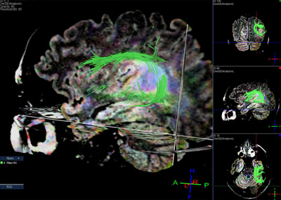
Fig 3. This is a sagittal T2-FLAIR image of the same patient seen in figure 1. The arcuate fasciculus (green fibers) is seen to wrap around the T2-FLAIR hyperintense mass. The arcuate fasciculus connects regions of the brain involved in language. This again indicates that in this patient resection of the mass would likely cause language deficits.