
Pathology
Cancer research often begins with tissue—a tumor sample, a blood draw, a slide. The Pathology Shared Resource can supply the specimens and help turn them into data.
New Project Consultation
Starting a new project involving one or more Shared Resources, or interested in learning more about available services? Contact Dr. Kate Hyde to schedule a consultation or complete the consultation form in Stratocore PPMS.
Request Service
Use Stratocore PPMS to request core facility services and manage scheduling and billing.
Pricing Benefits
Subsidized pricing is available to Fred & Pamela Buffett Cancer Center members. If you haven't received an email about subsidized pricing, please get in touch with cancer center administration to see if you qualify.Contact: buffettcancercenter@unmc.edu
If you conduct cancer-related research at UNMC and are not yet a member of a Cancer Center research program, visit the Fred & Pamela Buffett Cancer Center Membership page to learn more about becoming a member.
Institutional Biospecimen Bank
The Institutional Biospecimen Bank (IBB) is the University of Nebraska Medical Center’s central source for human biospecimens. It collects, processes, stores, and distributes human tissue and blood samples donated by patients who have consented to their use in research, along with the associated clinical data. | Pricing Information
Services
Getting Specimens
-
Tumor and normal tissue: formalin-fixed paraffin-embedded (FFPE), snap frozen, viably frozen, and fresh
-
De-identified serum, plasma, and whole blood
-
Sterile fresh tissue collected directly in the operating room
-
Retrieval of archived patient tissue
Processing Specimens
-
DNA, RNA, and protein extraction, using an automated system that works across blood, saliva, and tissue
-
Peripheral blood mononuclear cell (PBMC) isolation, performed by hand
-
Short-term tissue culture
Requesting Specimens
Requests are submitted through Stratocore PPMS.
Tissue Sciences Facility
The Tissue Sciences Facility provides histology, immunohistochemistry, and digital pathology services for human and animal tissue. | Pricing Information
Services
Preparing Tissue
- Tissue processing, paraffin embedding, and sectioning
- Frozen sectioning (cryosectioning)
Staining
- Routine and special stains for paraffin and frozen samples
- Immunohistochemistry, including multiplex immunohistochemistry and immunofluorescence—labeling several markers on a single slide to see how different cell types sit in relation to one another
- Antibody optimization and assay development
Scaling Up and Digitizing
- Tissue microarray construction: placing many small tissue samples on a single slide so that dozens or hundreds of specimens can be stained and compared under identical conditions
- Whole-slide digital scanning (brightfield), producing images you can analyze quantitatively
Support
- Pathology consultation and experimental design guidance
- Training on specialized histology procedures and equipment
- Self-service access to instrumentation after training
Instruments
- Ventana Discovery Ultra: Automated immunohistochemistry, including multiplex staining
- Leica CM1860 Cryostat: Sectioning of frozen tissue
- Tissue-Tek Prisma/Glas : Automated stainer and coverslipper for consistent, high-volume routine staining
- Leica Aperio CS2: Brightfield whole-slide scanner
- Hamamatsu NanoZoomer S360: High-throughput digital slide scanner with an 80x optical objective
- Ultra Freezers: These freezers are used to store a growing biobank of over 5,000 viably frozen tumor samples and over 10,000 snap-frozen tumor samples.
Submitting Samples
Place orders through Stratocore PPMS. Standard turnaround time is five to ten business days depending on complexity. Antibody optimization, tissue microarray construction, and image analysis are quoted separately. Orders are accepted Monday through Friday, 9:00 a.m. – 3:45 p.m.
Please contact the facility before starting a project, as decisions about fixation and embedding are difficult to undo later, and a short conversation upfront prevents most delays.
Citing Shared Resources
Researchers using the Fred & Pamela Buffett Cancer Center Shared Resources for publications, presentations or other scholarly outputs are required to acknowledge the use of them. Proper acknowledgment helps support the continued availability and funding of these cores.
Please acknowledge the Cancer Center Support Grant (P30 CA036727) in all publications and abstracts that have used these services.
Suggested language: Research reported in this publication was supported by the Pathology Shared Resource and the National Cancer Institute under award number P30 CA036727. The content is solely the responsibility of the authors and does not necessarily represent the official views of the National Cancer Institute.
Co-Directors
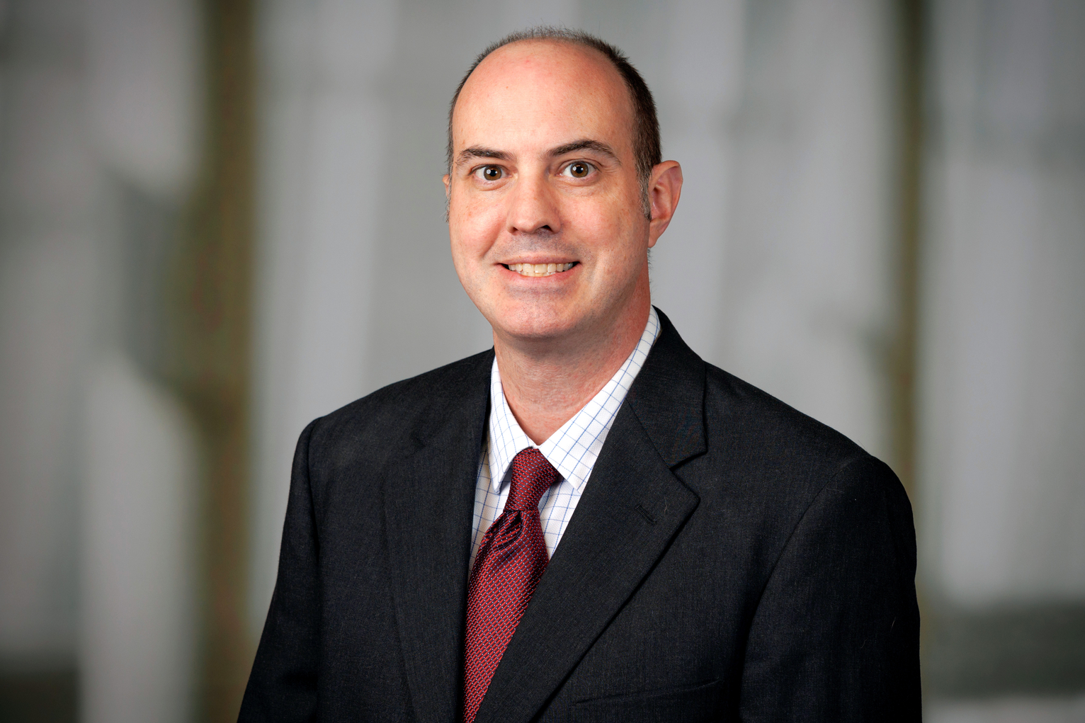
Kurt W. Fisher, MD, PhD
Co-Director, Pathology Shared Resource
Director, Institutional Biospecimen Bank
BCC SRT 5.12.431 | Send Email | 402-559-5086 | 8:00 a.m. – 5:00 p.m.
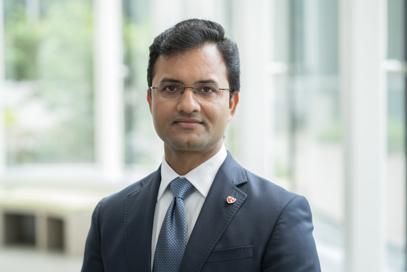
Dinesh Pradhan, MD
Co-Director, Pathology Shared Resource
Director, Tissue Sciences Facility
DRC1 1052 | Send Email | 402-559-5123 | 9:00 a.m. – 3:00 p.m.