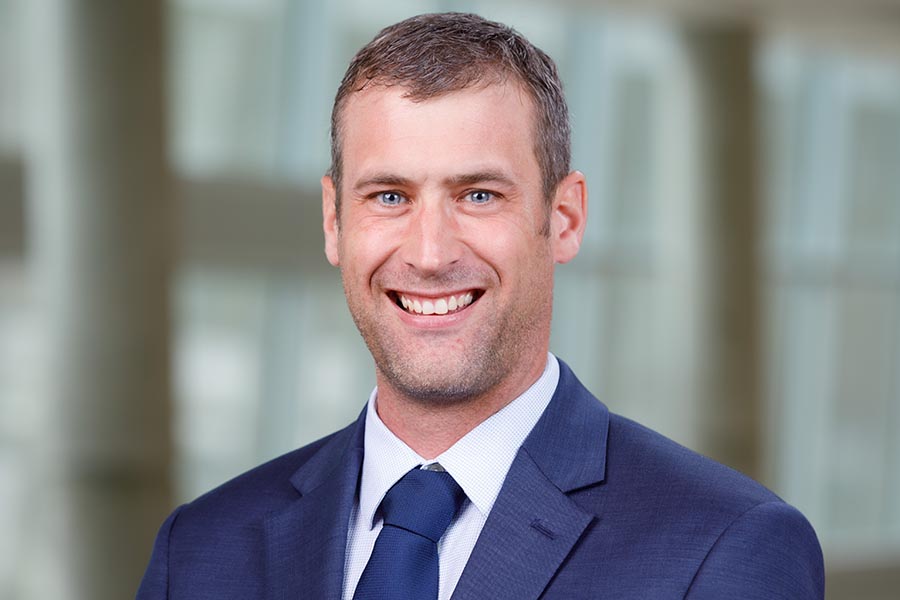
Bryan T. Hackfort, PhD
Assistant Professor, UNMC Department of Cellular and Integrative Physiology
Director, three research cores

- The Echocardiography Imaging Facility has two Sonosite, Fujifilm Vevo 3100s (one with Lazr-x for photoacoustic imaging) and a Revvity, Sonovol Vega for high through put ultrasound imaging.
- The Animal Physiology Core of the Center for Heart and Vascular Research has three surgical stations for independent surgeries. Each station has a heated surgical stage, ECG monitoring, microscope, and a ventilator, if needed.
- The Bioassay Core of the Center for Heart and Vascular Research has many instruments to assist in the processing of tissue and/or samples for a variety of research including immunoassays, western blots, RNA processing and cell sorting for flow cytometry.
Education
- Phd, Creighton University, Omaha, 2014
Research
- Publications, PubMed
Department of Cellular and Integrative Physiology
College of Medicine
University of Nebraska Medical Center
985850 Nebraska Medical Center
Omaha, NE 68198-5850