Modeling Optic Nerve Regeneration
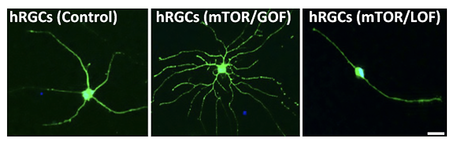
The mTOR pathway is involved in determining the growth and complexity of neurites, thus identifying it as a potential target for axon regeneration (image: Pooja Teotia)
The degeneration of the optic nerve in glaucoma leads to irreversible blindness as the connection between the retina and higher center of the brain is lost. We are testing our hypothesis that recapitulation of developmentally relevant gene expression and pathways, particularly those underlying neurites growth and guidance, promote optic nerve regeneration, using the microfluidic model of optic nerve regeneration developed in our lab (Teotia et al., 2019 Development). We have demonstrated that the mTOR pathway is developmentally recruited for the structural and functional differentiation of human RGCs. Particularly, it is involved in determining the growth and complexity of neurites, thus identifying it as a potential target for axon regeneration (upper panel). Examination of hRGC axon regeneration in microfluidic devices revealed that the number of regenerated axons and their length were significantly higher in the mTOR gain-of-function (GOF) group versus mTOR loss-of-function (LOF) and untreated groups, following axotomy (lower panel). Further, we present evidence that the mTOR pathway mediates the regenerative effects of other developmentally relevant pathways recruited by IGF-1 and guidance molecule such as Netrin-1 (Subramani et al., 2023 Stem Cells). These developmentally relevant pathways have been implicated in the regeneration of optic nerve in animal models. Hence, our observations show that the role of these pathways in optic nerve regeneration is evolutionarily conserved, suggesting their clinical relevance in glaucomatous degeneration. Currently, we are examining how the mTOR pathway integrate diverse regeneration promoting signals.
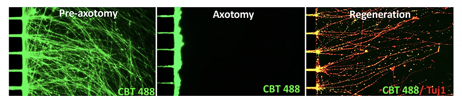
Examination of hRGC axon regeneration in microfluidic devices revealed that the number of regenerated axons and their length were significantly higher in the mTOR gain-of-function (GOF) group versus mTOR loss-of-function (LOF) and untreated groups, following axotomy. (image: Murali Subramani)