UNMC_Acronym_Vert_sm_4c
University of Nebraska Medical Center

Intravital Imaging Core

Core Director

Padmashri Ragunathan, PhD

Assistant Professor, Developmental Neuroscience, UNMC Department of Neurological Sciences

Padmashri Ragunathan, PhD

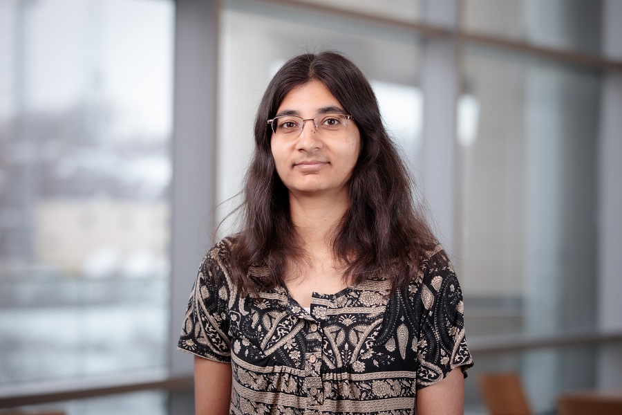
Dr. Ragunathan is an associate professor in the Department of Neurological Sciences at the University of Nebraska Medical Center. Dr. Ragunathan received her postdoctoral training at University College London and University of Nebraska Medical Center. Her research focuses on understanding neuronal and astrocyte alterations during development and with learning in mouse models of neurodevelopmental disorders using electrophysiological, in vivo imaging and behavioral approaches.

Did you use data generated at the UNMC Intravital Imaging (MITI) Core in your grant proposal or scholarly publication? Please cite or acknowledge the Intravital Imaging Core in your publications by using its unique Research Resource Identifier (RRID): RRID:SCR_022478.

Core Equipment

Contact Dr. Ragunathan for more information or for scheduling.

Coming Soon!
Tissues are inherently three dimensional in nature, which makes imaging intact tissues a necessity for a more complete study in the relationship between structure and function and the system-level study of cellular mechanisms. The Miltenyi Biotec (previously La Vision) Ultramicroscope II Light Sheet fluorescence microscope (LSFM) extends your existing fluorescent imaging into true 3D, large-scale volumetric imaging the nervous system. Optimized volumetric imaging requires removal of light bending cellular components (mostly lipids) through a process called optical clearing.
 
Cost: $40/h for assisted microscopy. Users accruing significant experience and training during assisted imaging sessions are eligible for unassisted imaging with technological assistance available ($30/h) and/or unassisted imaging without technological assistance ($20/h). Unassisted imaging options are approved at the discretion of Dr. Jensen-Smith.
Tissue clearing is important for 3D imaging of tissues at single-cell resolutions. The X-CLARITY™ system simplifies and accelerates each step of the tissue clearing process using CLARITY (Clear Lipid-exchanged Acrylamide-hybridized Rigid Imaging / Immunostaining /in situ-hybridization-compatible Tissue hYdrogel). Native cytoarchitecture remains intact and endogenous fluorescence proteins are preserved for robust fluorescence imaging downstream. For LSFM imaging, samples should be optically cleared (X-clarity), labeled with antibodies, then equilibrated in refractive index (RI) matched mounting solutions for imaging.
 
Cost: $100/run. Each run includes hydrogel embedding and subsequent clearing. Researchers are responsible for subsequent antibody labeling (if applicable) and refractive index matching prior to imaging. Large sample may require extended clearing ($150/ long run).
IMARIS is the world’s leading Interactive Microscopy Image Analysis software focused on 3D and 4D imaging. The complete IMARIS for Core Facilities software package with focused image analysis, segmentation, and reconstruction options for cancer and neuroscience researchers, cell and developmental biologists is available for use. IMARIS can be used for any number of imaging studies but is anticipated to be particularly relevant for researchers using the Ultramicroscope II light sheet fluorescence microscopes.
 
Cost: $5/h unassisted, $40/h assisted.