Our department has been actively engaged in telepathology and digital whole slide imaging (WSI) efforts for the past several years. These efforts include education, research, and emerging applications for clinical workflow.
Digital Pathology has revolutionized teaching.
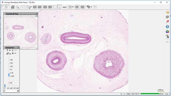
Learning is not confined to the classroom. Virtual microscopy has replaced the use of optical microscopes in education. Microscopic WSI is integrated into pathology courses for medical students, residents and allied pathology schools (e.g., cytotechnology). Digital images are available to a large number of students with full access anywhere at any time. Compared to glass slides the quality of the image can also be indefinitely maintained and the risk of breakage is eliminated. UNMC uses PathXL as part of the educational experience. This web based program allows educators to create clinical vignettes with associated attached radiographic, gross, and histopathology images. Medical students can log on and work through these virtual cases where and when they desire either on or off campus. Digital pathology is a powerful educational tool that replaces the traditional standard methods of teaching and learning pathology. WSI has been used in our department for the creation of pathology resident teaching sets, as well as interdepartmental tumor board conferences.
Digital Pathology in Research
Currently, our department offers digital WSI services to UNMC researchers campus wide using a Leica Biosystems CS2 scanner located within our Tissue Science Facility (TSF). Investigators studying animal models can submit glass slides of various organs for scanning and in turn receive WSI via a cloud based server. These researchers also have access to an image analysis program located in the TSF if desired for quantitation, etc.
Digital Pathology/Telepathology is enhancing Clinical Workflow
We have begun a new phase in our diagnostic services by incorporating telepathology as a key component. Currently, our pathologists use remote live microscopy, using the Mikroscan system, for interpretation of frozen sections at both Bellevue Medical Center and Village Pointe. This allows the pathologist to remain on campus and improve overall workflow efficiency. Soon, remote frozen section interpretation will be further improved with the addition of Leica Biosystems CS2 WSI scanners. With a scanner located in the main frozen room on campus, pathologists will be able to read frozen sections such as donor organ samples from home during non-business hours. The workflow for assessing adequacy of cytology specimens and kidney biopsies has also been improved by utilizing iPad technology in combination with an in-house designed microscope adaptor. This allows the pathologist to visualize both cytology smears and kidney cores remotely both on campus and at regional medical centers. Implementation of digital pathology will also provide second opinion expertise when needed to our pathologists who are staffing remote locations, such as Kearney Regional Medical Center. In addition, the department is excited to begin using digital WSI for primary diagnosis including full integration with our laboratory information system. For this purpose, the department has acquired a Leica Biosystems GT450, their most advanced high throughput scanner.
For further information on this initiative, please contact Kirk Foster, MD 402-559-4186.