 |
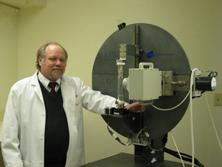
Lee Mosley, Ph.D., manages the SPECT. |
The Single Photon Emission Computed Tomography (SPECT) allows researchers to visualize functional information about a subject’s specific organ or body system.
“The use of SPECT technology is limited only by the imagination and the appropriate probe for your application,” said Lee Mosley, Ph.D., assistant professor in the Department of Pharmacology and Experimental Neuroscience at UNMC.
Dr. Mosley manages the SPECT with Howard Gendelman, M.D., chairman of the Department of Pharmacology and Experimental Neuroscience and director of the Center for Neurovirology and Neurodegenerative Disorders.
Although, a long-term goal is to centralize UNMC’s imaging equipment into an integrated core, Drs. Gendelman and Mosley simply want investigators to know the SPECT is now available and assess its need in support of the research community.
“Imaging is emerging as a huge need on campus,” Dr. Gendelman said. “This technology is significant and we have some facilities that are under and over utilized. It’s important for the research community to know what we have now and how it can best be used for the good of all.”
In many ways the collaboration already has begun.
“There is a growing need for imaging,” said Paula Turpen, Ph.D., director of Research Resources at UNMC. “Technology is being developed all the time that allows us to acquire new information. New labeling reagents are developed and combined with new imaging technologies to improve the resolution of what we can see in a living organism.”
The SPECT — best utilized as a tool for non-invasive or non-terminal procedures that need to be followed over a period of time — primarily has been used to address questions involving inflammation, neurodegeneration and tracking of immune cells associated with regulation of inflammatory responses and neuroprotection.
That, however, is changing. “In collaboration with other investigators, we have utilized the SPECT for a very diverse range of research endeavors,” Dr. Mosley said.
Investigators in UNMC’s Radiation Oncology Department have used the SPECT to assess the increased uptake by tumors of a tumor specific antibody in an effort to enhance radioimmunotherapeutic agents in cancer. A pharmaceutical sciences researcher has used the SPECT to assess the biodistribution of drugs in rheumatoid arthritis and osteoporosis. In another collaborative study, researchers studied the delivery of nanoformulated compounds across the blood brain barrier.
With SPECT, internal radiation is administered via a pharmaceutical that is labeled with a radioactive isotope, which is injected, ingested or inhaled. The radioactive isotope decays, resulting in the emission of gamma rays and a 3-dimensional picture of what’s happening inside the body of the experimental animal. The animals are anesthetized and the probes or live cells are tracked for the duration of the experiment. A series of 64 images are acquired at equiangular points as the animal is rotated 360° and reconstructed into a single 3-dimensional image.
SPECT imaging can be used to diagnose a multitude of medical conditions including heart disease, certain forms of infection, the spread of cancer, and bone and thyroid diseases. Brain SPECT studies help in the diagnosis of brain trauma, dementia, atypical or unresponsive mood disorders, strokes, seizures, the impact of drug abuse on brain function, complex forms of Attention Deficit Disorder, and atypical or aggressive behaviors.
In animal models, such non-invasive assays as SPECT provide an added benefit, Dr. Mosley said. With the technology the same animals may be longitudinally followed over time, he said, which eliminates the need for separate animal groups at each time point, reduces large cohorts of animals for each experimental group and reduces the inter-animal variability.
In addition, Dr. Mosley said, similar imaging systems may be used to gauge therapeutic efficacy in clinical studies. “SPECT, PET (Positron Emission Tomography) and MRI (magnetic resonance imaging) technologies are the techniques of choice for objective assessment of patients with neurodegenerative disorders and therapeutic outcomes,” Dr. Mosley said. “These technologies should provide a much-needed dimension to not only preclinical studies, but also to translational endeavors.”
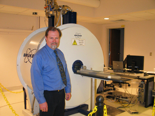 |
Michael Boska, Ph.D., manages the MRI facility. |
Researchers also are combining imaging techniques. Linking MRI and SPECT provides investigators a more complete picture, Dr. Mosley said, as the imaging techniques provide complementary information.
The MRI scanner can pick out a tiny point inside the patient’s body and essentially ask: “What type of tissue are you?” The point might be a cube that is a millimeter on each side. The MRI system goes through the experimental animal’s body point by point, building up a 2-D or 3-D map of tissue types. It then integrates all of this information together to create its multi-dimensional models.
“The combination of SPECT and MRI technologies will provide the University community higher levels and greater dimensions of non-invasive small animal real-time measurements of benefit to basic and translational research efforts,” Dr. Gendelman said.
If you are interested in using the SPECT, contact Dr. Mosley at 559-2510. If you are interested in using the MRI technology, contact Dr. Boska at 559-3138. Read an overview on SPECT at UNMC.