A boost for women's health
At UNMC, iEXCEL medical animation is giving students and patients greater understanding of the female pelvis and how even small changes in the anatomy of the pelvic floor can have a major impact on a woman's quality of life.
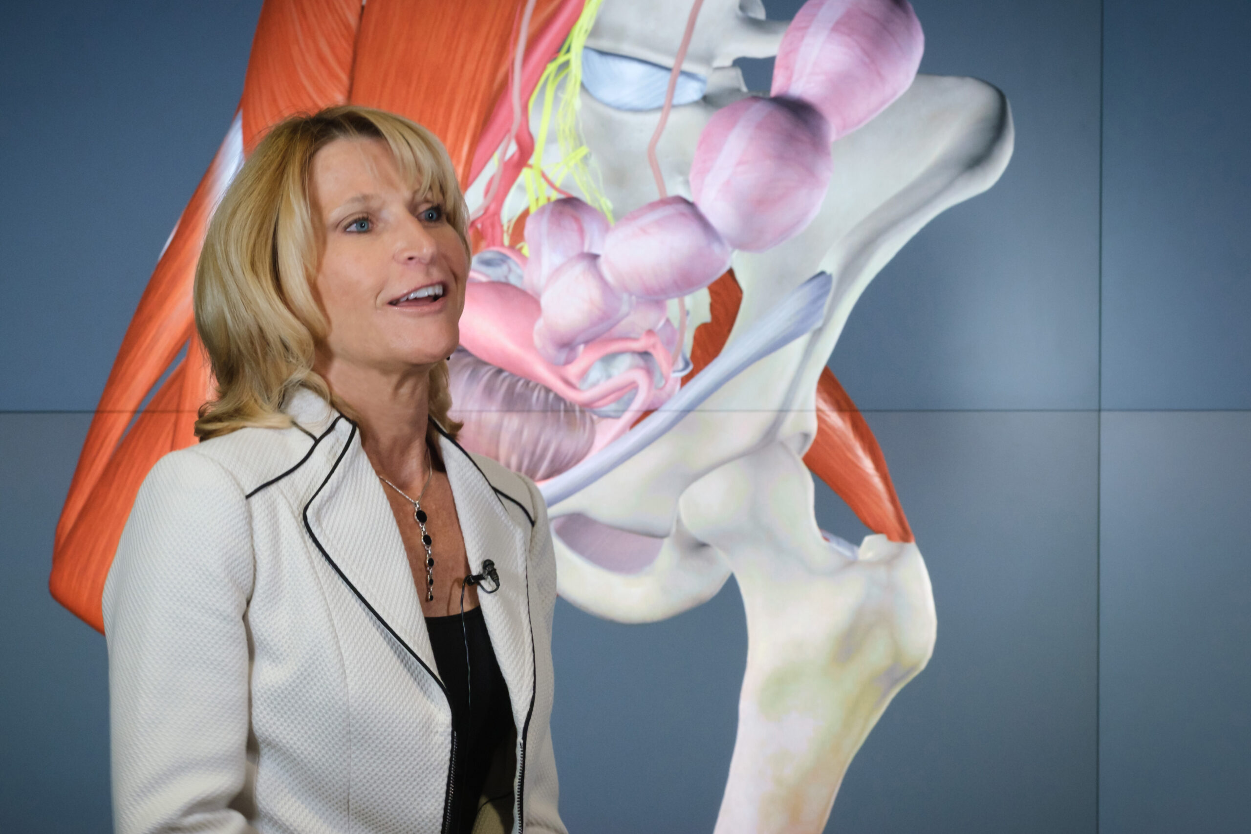
Dr. Jennifer Cera demonstrates the 3D model of the musculoskeletal system of the female pelvis created by the iEXCEL visualization team at UNMC. Photography by Kent Sievers/UNMC Strategic Communications.
Jennifer Cera, DNP, walked into a brain, and it blew her mind.
The brain was a model, of course, during an iEXCEL open house, in 2019. iEXCEL is UNMC’s transformative program for health care education, training and research, which uses simulation, advanced technology, interprofessional collaboration and experiential learning. The open house was an initial attempt to give faculty and others a glimpse of the program’s capabilities and possibilities. But, Dr. Cera understood immediately.
“It was like a light bulb just came on,” said the assistant professor and co-coordinator of the Women’s Health Nurse Practitioner Program at UNMC College of Nursing. “I knew instantaneously what this could mean for my students and for my patients.”
Dr. Cera explains how she came to request the 3D interactive teaching model. Videography by Rich Watson/UNMC Strategic Communications
Dr. Cera specializes in women’s health, specifically pelvic floor health. She’s passionate about her specialty, a quality-of-life issue for so many women, she says. Because pelvic-floor issues, often following childbirth, are so common, yet these issues still are not often discussed.
“All of these things are so preventable,” she said. And treatable, yet not enough education is imparted to women at all phases of their life.
“Women will tolerate some pretty severe conditions,” said Julie Mayberger, who studied under Dr. Cera and recently graduated from UNMC with a Doctor of Nursing Practice (DNP).
If only Dr. Cera could show people – students, future practitioners of every profession; and, yes, patients, too – what was going on inside.
She had been hunting for a model, a good model, for years. Something that would improve students’ mastery of what was happening, spatially, within the pelvis, with emphasis on pelvic organ prolapse. Something that was an impactful experience to improve comprehension of how specific patient complaints correlate to the changes in the female pelvic anatomy.
The iEXCEL visualization staff was eager for the challenge.
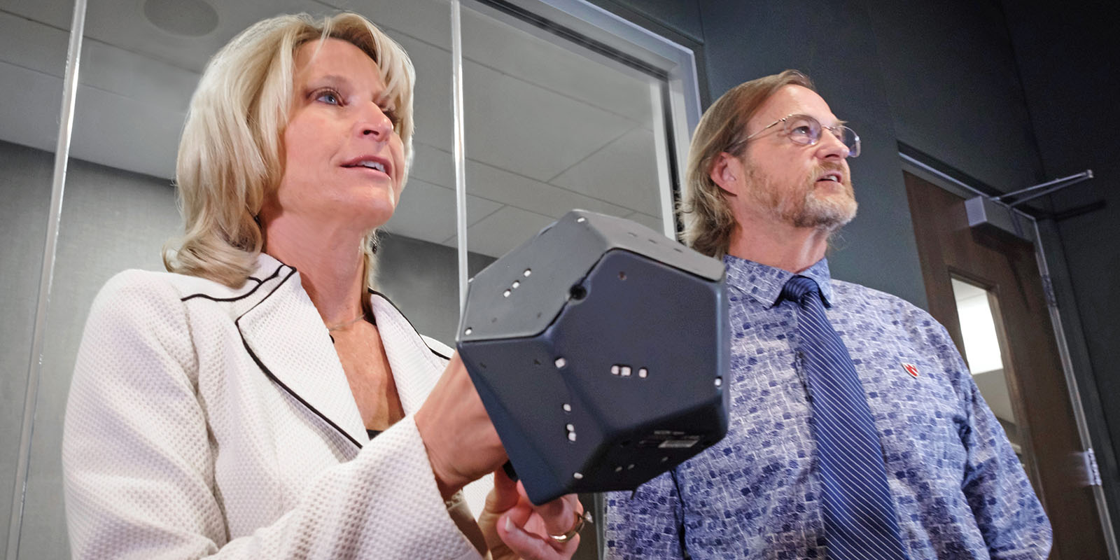
Dr. Jennifer Cera and Bill Glass collaborated on the pelvic model's versatility and adaptability. The content is designed to be both student- and patient-friendly.
“Our overall goal is to build out an entire body,” said Bill Glass, iEXCEL’s artistic director. “We like to start with CT and MRI data, so when Dr. Cera came to us with this project, it was something we were hoping we could take on.”
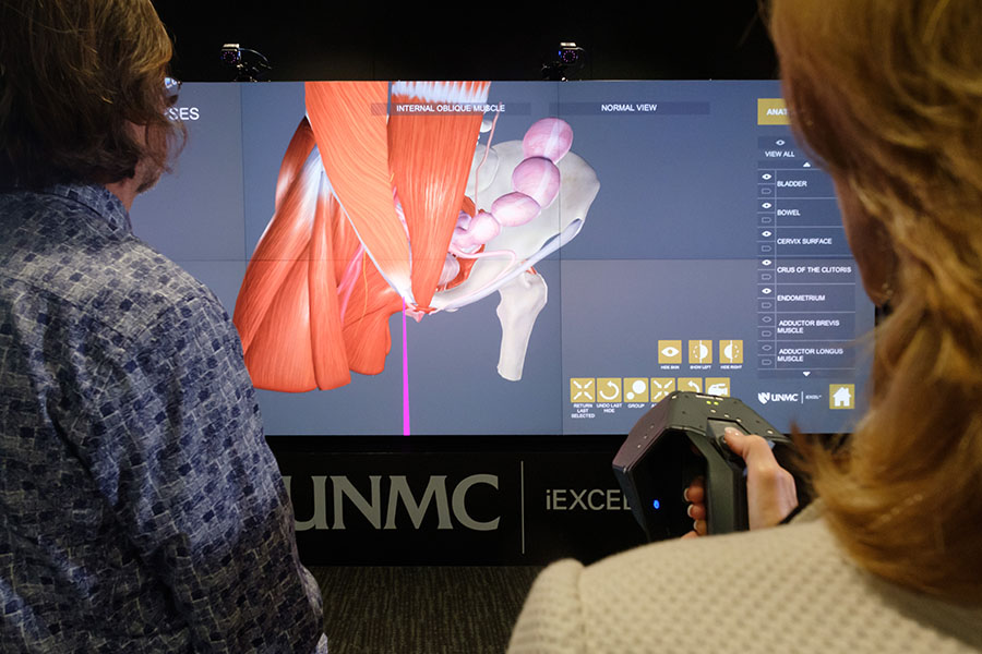
iEXCEL took on the project in 2019, working on it off and on. It probably involved a total of six months of work over those two years. The result: A 3D virtual reality pelvic model for three learning platforms: iWall, CadWall and zSpace.
“I loved the zSpace because I had control over it,” said Dr. Mayberger, who was one of the first students to use the model in a focus-group pilot study. “I could actually grab pieces of anatomy and move them around.”
Like never before, it’s possible to see the pelvic floor’s urogenital, musculoskeletal and neurovascular systems.
“You can see how prolapses can affect adjacent organs and why patients report the symptoms they do. How it is affecting the other organ systems,” Dr. Mayberger said.
Seeing this anatomy on a piece of paper doesn’t do it justice, she said. A 2D model can only take your understanding so far. The only thing comparable would be taking part in a surgery, Dr. Mayberger said.
“Being able to become immersed in it, it’s powerful,” Dr. Cera said.
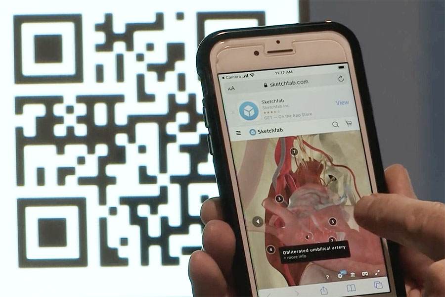
A total of five QR codes take users to iEXCEL-generated medical animations of the pelvic floor. The animations are interactive, showing various components of the female anatomy.
Dr. Cera currently teaches nursing students. But, her goal is to use this tool to reach everyone who treats women. Medical students. Orthopedic surgeons. Allied health professionals.
“I find a lot of happiness in not only improving a patient’s quality of life and empowering them with the education,” Dr. Cera said, “but in teaching students that they can also go out and provide this care as well.
“I looked for years to find a simulation mannequin or any type of model that would adequately reflect the changes in structure and function due to pelvic prolapse, but there weren’t any. So, when I was able to be immersed within the brain at iEXCEL’s open house, I knew this was it,” Dr Cera said.
With this model, clinicians will know to look for, “Things they can’t necessarily tell you,” Dr. Mayberger said.
“This has been a labor of love to provide the content to as many students as well as patients as I can,” Dr. Cera said. “Developing a 3D virtual reality pelvic model where students are immersed within the content, so they can really see and comprehend pelvic anatomy and pelvic organ prolapse, will spill over to their clinical expertise and mastery.
“And without iEXCEL, we could have never done this.”
Try Iit Out
Interact with the technology for yourself. Scan this QR code with your smartphone camera to see the iEXCEL 3D model of the innervation supply of the female pelvis. The visualizations help students and patients more clearly understand the female anatomy.


Bill Glass explains the iEXCEL approach to medical animation.