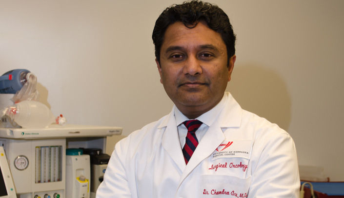
When Chandra Are, M.B.B.S., arrived at UNMC in 2007, the anatomy lab had recently become an early adopter of the lightly embalmed process for cadavers. Dr. Are saw an opportunity for collaboration to enhance resident training using this updated method — and within two years he was conducting 20 sessions a year.
What is “lightly embalmed”
In traditionally embalmed cadavers preserved with formaldehyde, the tissues become hard, tough, and leathery. Internal organs and structures are difficult to distinguish because all of the body tissues become shades of gray. The blood also solidifies, so the bleeding that takes place in surgery is not replicated. The normal pliability and varied coloration of living tissue is lost. These differences between living tissues and traditionally embalmed cadavers limit somewhat the types of procedures and lessons that can be done.
However, the tissue of lightly embalmed cadavers is much more similar to living tissue and can therefore be a better model for learning surgical techniques. In a lightly embalmed cadaver, the tissues more closely resemble in look and feel the living tissues surgeons work on in the operating room. Lightly embalmed cadavers are so similar to living tissues, Dr. Are said, that when he shows images of the lightly embalmed cadaver procedures to residency applicants, they often cannot immediately distinguish them from images taken during actual surgeries.
“Lightly embalmed cadavers are as close to real life as possible,” he said, and therefore are an invaluable tool for resident training.
“A new training program needs a champion,” said Dr. Are, who guided the program through its early development.He recalled one time when residents were scheduled to remove a cadaver’s appendix, only to find it was already gone.
Once the program was running smoothly, Dr. Are reached out for collaborators to continue growing the program, inviting other surgical faculty members to join in, and 15 to 20 faculty have now participated.
The operating room can be a stressful setting, but the cadaver lab provides a low stress environment for discussion. Currently, residents practice as many as 25 to 50 common procedures annually, with increasing difficulty as they advance in skill. Later, when operating on patients, the residents mention their experience working with the cadavers and how they can apply the skills they developed.
“This surgical training allows residents to walk before they run,” Dr. Are said, because the lightly embalmed cadavers more realistically mimic an actual surgery (see sidebar).
The program provides a very cost-effective method of teaching surgical skills and anatomy, Dr. Are said, and surgical residents consistently give the program the highest rating among their training activities.
The open surgical skills training program using lightly embalmed cadavers sets the UNMC surgical program apart from programs at other universities, Dr. Are said. Residents interviewing at UNMC appreciate the unique program, which helps to recruit top residents to Nebraska. Dr. Are and his colleagues have published research on the program’s success in the hopes of setting an example for other academic medical centers.
“The program has already been shown to be feasible, acceptable, and appreciated,” Dr. Are said, so he has expanded the program to include first-year medical students, giving them exposure to surgery earlier in their training.
In the future, he hopes to expand the program further to include more opportunities for interprofessional education through participation by other disciplines, with the program serving as a basis for collaborations in training and research across departments.
Dr. Are said he plans to use detailed metrics to further assess the program and continue improving it and that he hopes to find additional funding support to move the program to the next level.