 Jon Henning and four other medical students huddle around the waist-high table of their cadaver in search of the brachiocephalic vein.
In the past, they would have flipped through a chunky anatomy atlas to find the vessel. Now, guiding their dissection is a 50-inch screen that projects the precise location of the vein in the upper chest.
It’s a new world inside UNMC gross anatomy, which has moved from a strong reliance on textbooks and anatomy atlases to online images.
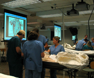
Today, medical, physician assistant and physical therapy students at UNMC are studying gross anatomy in a newly renovated lab with computers at each cadaver station. Instead of viewing anatomical images in a textbook, students view enlarged images online via 29 video projectors and pull-down screens.
At the foot or head of each cadaver station, a 50-inch projection screen hangs from the ceiling to enhance the students’ 10-week introduction of the structural organization of the human body.
 “The renovations are amazing,” said Todd Lovgren, a fourth-year medical student. “Nebraska’s anatomy lab took a big step into the 21st century.”
In the past, students spent precious lab time paging through a cadaver dissection guide developed by UNMC’s Robert Binhammer, Ph.D., and a hefty, commercial anatomy atlas. Today, the contents of the soft-covered dissection guide are available on the Web, allowing students to find anatomical terms, definitions and images with the click of a mouse. The key anatomical words in the guide are linked to nearly 700 images in Frank Netter’s thick human anatomy atlas.
“Displaying the images so everyone can see is a real benefit,” said Gordon Todd, Ph.D., gross anatomy instructor. “Anything you can do to increase the students’ efficiency in learning the material is beneficial.”
Henning agrees, glancing toward the projection screen. “(The atlas) is pretty voluminous so it takes a long time to find the exact slide. The links allow us to go directly to the slide.”
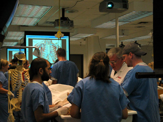
A mouse at each cadaver station gives students the ability to project the information on the large screens for all to see, rather than crowding together to view the smaller, textbook images. Being able to point directly to structures on the screens enhances the small, group teaching environment.
“Reviewing important structures in the gross anatomy lab has become more productive than before,” said Ben Solomon, a second-year medical student. “Students can spend less time flipping through Netter’s Atlas of Human Anatomy and more time focusing on the cadaver. It also makes the lab a little more interactive since all five students in the lab group can easily view the same picture on the screen instead of crowding around one book.”
Andrew Livingston, a second-year medical student and gross anatomy tutor for first-year medical students, has seen the benefits of the project. “This method of combining virtual gross anatomy with the time-honored tradition of cadaver dissection well illustrates that the latest technology can be combined with medical education,” he said.
The Olney Family Foundation, the Alumni Class of 1953 and the UNMC College of Medicine funded the gross anatomy renovations.
As a gross anatomy tutor for first-year medical students, Livingston said he benefits from the advantages. “I can point to the projected slides and immediately reinforce what we are reviewing on the cadaver,” he said. “The advancements in the gross lab save time and leave more time for dissection.”