Researchers in the magnetoencephalography (MEG) laboratory currently have ongoing research studies for healthy adults, as well as HIV-infected patients, and are seeking study participants.
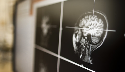
MEG is a brain imaging device that allows researchers to see the brain in action. It measures the magnetic fields that are naturally produced by the brain cells. These magnetic fields are created when the brain cells communicate with each other using electrical signals and thus, the MEG can analyze how the brain solves problems.
UNMC/Nebraska Medicine started using MEG in 2009 and was one of the first institutions to use the device in the United States. There are currently some 30 institutions that utilize MEG and that number is expected to rise as researchers learn its value.
“MEG allows us to see the brain in action, which no other method can do,” said Tony Wilson, Ph.D., associate professor of pharmacology/experimental neuroscience & neurological sciences, vice chair of basic/translational research, and scientific director of the MEG Laboratory. “The average adult reads three words per second, and the MEG is able to see how the brain does this millisecond by millisecond. It is amazing.”
The ability to see these results has led to a better understanding of many diseases such as Parkinson’s, autism, Alzheimer’s, schizophrenia and many others.
Learning how these diseases affect the brain may help to improve outcomes for people affected by them. If you or someone you know is interested in participating, please contact Mackenzie Mills or Nichole Knott at 402-552-6444 or email Mackenzie.mills@unmc.edu for more information. Participants will be compensated for their time. (IRB #225-14-EP.)