 |
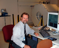
Michael Boska, Ph.D., with the MRI unit he used for his research. |
“This is an exciting new development,” said Michael Boska, Ph.D., associate professor in the UNMC Department of Radiology and principal investigator on the study. “Typically, Parkinson’s disease is not diagnosed in humans until about 50 percent of the brain neurons that control body movement are damaged. By the time it is diagnosed, it is too late to reverse the damage. With this new MRI technique, we are now able to detect Parkinson’s disease in rodents when as few as 25 percent of the brain neurons that control body movement are damaged.”
The study bridged divergent scientific disciplines, representing collaborative efforts between investigators in the departments of radiology and pharmacology and the UNMC Center for Neurovirology and Neurodegenerative Disorders (CNND).
Other principal scientists contributing to the project included Howard Gendelman, M.D., director of the CNND and chairman of the UNMC Department of Pharmacology, and R. Lee Mosley, Ph.D., assistant professor of pharmacology and chief of the CNND’s movement disorders program.
“We’re optimistic that with more studies we will be able to make diagnosis even earlier,” Dr. Mosley said. “Earlier detection would make a huge difference in treatment of the disease. If this same technology can be translated to humans, this would be a significant development.”
Parkinson’s disease is a chronic, debilitating disease without a cure. After Alzheimer’s disease, Parkinson’s disease is the most common neurodegenerative disease. It affects 1 in 100 people over the age of 60, with the average age of onset being 60 years. Approximately 500,000 people in the United States have Parkinson’s disease with as many as 50,000 new cases diagnosed each year.
The disease occurs equally in men and women and can also affect younger people. Young-onset Parkinson’s disease (onset at age 40 or younger) is estimated to occur in 5 to 10 percent of patients with Parkinson’s disease. The disease is characterized by four major features: tremor of a limb; slowness of movement; rigidity of the limbs or trunk; and poor balance.
When at least two of these symptoms are present, and especially if they are more evident on one side than the other, a diagnosis is typically made. Symptoms typically begin on one side of the body and spread over time to the other side. Both younger and older patients may experience initial symptoms for a year or more before seeking medical evaluation.
The new study used magnetic resonance spectroscopic imaging (MRSI), a variation of magnetic resonance imaging (MRI), which is available in most hospitals. MRSI, which can be done in humans, uses a standard MRI unit equipped with custom software.
The study used a MRI research unit with a magnetic field strength of 7 Tesla. The unit is located at The Nebraska Medical Center, the teaching hospital for UNMC. Higher field systems of MRI improve the signal quality and can provide detailed chemical composition from tissue volumes the size of a pinhead.
Human clinical scanners are typically up to 1.5 Tesla. A new FDA-approved system with a field strength of 3 Tesla will be installed at The Nebraska Medical Center next fall in the Clinical Center of Excellence building now under construction. This new system will be an important tool for rapid scanning and early diagnosis of the brain, Dr. Boska said.
The MRSI technique allows scientists to make a chemical analysis of the brain without doing a biopsy. “By extracting the chemical make-up of the brain, we are able to make a precise determination of how much brain degeneration has occurred,” Dr. Boska said. “In essence, it’s a non-invasive biopsy of the brain tissue.” Previously, this sort of information could only be attained at autopsy or with a brain biopsy.
In June 2004, scientists at UNMC and Columbia University Medical Center in New York discovered a new vaccine approach that successfully prevents the death of brain cells in a mouse model of Parkinson’s disease. The findings appeared in the Proceedings of the National Academy of Sciences (PNAS) of the United States of America.
The MRSI technique was used to support the vaccine work in the June study. The new study represents a critical next step toward human vaccine testing, Dr. Boska said, as monitoring systems must be established to determine therapeutic responses in humans.
“We still have technical challenges that need to be overcome,” Dr. Gendelman said. “But, this is a significant step forward toward better diagnosis and treatment of Parkinson’s disease.”
“Dr. Boska’s use of MRSI is nothing short of cutting-edge for modeling disease in real-time,” said Harris Gelbard, M.D., Ph.D., professor, University of Rochester School of Medicine, and a collaborator with the CNND on a study looking at therapeutic interventions for HIV-dementia. “Most importantly, using MRSI has helped us determine whether neuroprotective drugs work in living animals, which is not always the case in cell culture studies. That Dr. Boska was able to utilize MRSI to study whether a very novel Parkinson’s vaccine approach is neuroprotective (i.e. therapeutic) represents a vanguard for neuroscience. I believe this is the future of how translational studies should be performed when modeling human disease.”
This work was supported, in part, by the Alan and Marcia Baer Foundation, the Fran and Louis Blumkin Foundation, Inc. and five grants from the National Institute of Neurological Disorders and Stroke and the National Institute for Mental Health, two of the institutes that make up the National Institutes of Health.