 If it weren’t for the research seed money that Tom Porter, M.D., received from UNMC in the mid-1990s, he’s not sure where the promising research on microbubbles might be.
For certain, the research wouldn’t be as advanced as to begin an NIH-funded, Phase I clinical trial looking at the promise of microbubbles in drug delivery, as will occur at UNMC in the next year.
And, without the initial seed money, it’s unlikely that scientists in 14 sites worldwide would be ready to research the value of microbubbles in helping physicians study the hearts of patients undergoing stress tests.
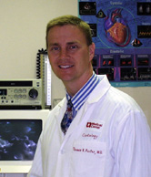
“I’m very thankful that the seed money allowed us to make a contribution in the cardiac care of patients,” said Dr. Porter, an associate professor of cardiology at UNMC. “And, we’re still trying to learn more.”
For his research work, Dr. Porter has been awarded the 2002 College of Medicine Joseph P. Gilmore Outstanding Investigator Award.
“Dr. Porter is a nationally recognized innovator in imaging of the heart,” said Ira Fox, M.D., associate dean for research and development within the UNMC College of Medicine. “The use of microbubble technology in cardiology could potentially have a major impact on how we evaluate and care for patients with heart disease.”
James O. Armitage, M.D., dean of the College of Medicine, and Dr. Gilmore, a UNMC professor emeritus, will present Dr. Porter with the award Wednesday at a 3 p.m. symposium in the Eppley Science Hall Amphitheater. Dr. Porter will then present the results of his research into the diagnostic and therapeutic applications of perfluorocarbon containing microbubbles. A brief reception will follow the symposium, which is open to the public.
|
Joseph Gilmore, Ph.D., was professor and chairman of the department of physiology from 1970 to 1987. He was funded by the National Institutes of Health for his entire career, supervised numerous graduate students and post-doctoral fellows, and published more than 200 scientific papers. The Joseph P. Gilmore Award was established by the department of physiology and biophysics upon Dr. Gilmore’s retirement in 1987 to recognize outstanding research contributions by a faculty member. |
A decade ago, hardly anyone would have thought that such a presentation could occur on microbubbles. Then filled with room-air gases, it was doubted that the microbubbles could reach the left side of the heart, where scientists could use them to study blood flow into the heart.
But Dr. Porter and his collaborative investigators thought that microbubbles could be altered to be usable in cardiac testing.
“The seed grant from UNMC allowed us to determine that if we changed the gas (inside the microbubble), they would reach the left side of the heart,” Dr. Porter said.
Initially, the testing, which took place on dogs, wasn’t too successful. During their experiments, though, Dr. Porter and his research group kept noticing that a bright light — almost like a starburst — would appear in the heart muscle immediately after they turned the ultrasound transducer on to the image of the heart. Eventually, they determined that their microbubbles were surviving until they were “hit” with the ultrasound. When this occurred, tremendous energy was given off, producing the contrast.
“Before this study, we hadn’t realized that the ultrasound was destroying the microbubbles,” Dr. Porter said.
What came about were two major medical advances: 1) By turning down the ultrasound power or delivering it intermittently, the patented microbubbles could remain intact and could be used to study the blood flow into the heart; and 2) that the microbubbles could be used for drug delivery. Researchers are studying ways in which drugs can be attached to microbubbles, delivered to a specific location, and then released in that area when ultrasound causes the bubbles to “bounce,” knocking the drugs off of the microbubbles.
“It really changed the way the everyone has viewed ultrasound,” Dr. Porter said. “That destruction process can be used beneficially. It’s still in its development, but this is a very promising, non-invasive technique.”
Dr. Porter said that one way in which the ultrasound technique will be studied therapeutically is in the area of breaking up clots that occur in clogged arteries. Scientists think that the destruction of microbubbles by ultrasound may erode the blood clot and restore flow in the blocked arteries.
For sure, using microbubbles in addition to the traditional echocardiogram will be further researched. An echocardiogram uses ultrasound to show physicians the thickness of the heart walls and to detect damage and disease. However, the test doesn’t show blood flow very well. Preliminary research indicates that the microbubbles can help physicians find heart disease that only the much more expensive nuclear imaging would be able to detect.
“Twenty percent of the stress tests in which we find a blockage to the heart using the microbubbles would have been called normal if only the traditional echocardiogram were used,” Dr. Porter said.