 |
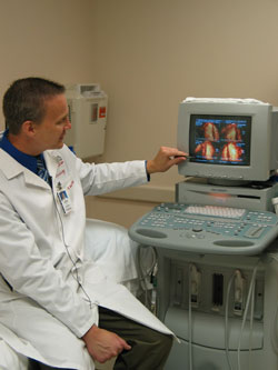
Thomas Porter, M.D., demonstrates the use of microbubbles to detect heart disease. |
Findings of a preliminary study — which was conducted by a research team led by Thomas Porter, M.D., professor in the UNMC Department of Internal Medicine — will be presented next week in Seattle during the 18th Annual Scientific Sessions of the American Society of Echocardiography. The abstract of the study was published recently in the Journal of the American Society of Echocardiography.
For the study, researchers evaluated a combination of traditional Dobutamine Stress Echocardiography — known as a “stress test” — and the new Real Time Perfusion Echocardiography — which is called “perfusion imaging” and involves the use of microbubbles. Diabetes patients with no symptoms or history of heart disease were used as subjects for the study because of their increased risk for heart disease.
“Almost 18 percent of the stress tests in which we found a blockage to the heart using the microbubbles would have been called normal if only the traditional echocardiogram were used,” Dr. Porter said. “The results show this microbubble contrast agent can detect blockages in the heart that may have gone undetected if conventional imaging had been used such as traditional stress echocardiograms or nuclear stress tests.”
When exposed to ultrasound, the microscopic bubbles are easily detected within the heart walls, enabling cardiologists to see the border and thickness of the heart walls, as well as the tiny blood vessels that feed the heart muscle. Traditional stress echocardiograms don’t detect blood flow in the heart muscle, he said.
A tiny amount of microbubbles are diluted with saline and administered to patients intravenously. The bubbles, smaller than the size of red blood cells, follow red blood cells through the heart and can identify areas of reduced or absent blood flow.
The study was conducted in 149 patients to determine the effectiveness and accuracy of perfusion imaging in detecting heart disease in patients with diabetes who had no symptoms of heart disease.
Blockages that would have gone undetected with traditional ultrasound were detected in 17 percent of the study participants, Dr. Porter said.
The patients, ranging in age from 26 to 84, also were followed for an average of 23 months afterwards to evaluate whether they had heart attacks or other heart problems. Three patients had non-fatal heart attacks during the follow-up period.
Omaha’s Linda Simpson was one of the 149 patients with diabetes to participate in the UNMC study. In 2005 she underwent a battery of medical tests, including a nuclear stress test, which found blockage. However, about six months later, she was having chest pain but this time enrolled in Dr. Porter’s study which revealed a blockage to another area of her heart.
“If they wouldn’t have found it, I probably would have had a heart attack. I owe my life to The Nebraska Medical Center. I’m glad the study was available,” Simpson said.
Dr. Porter and his colleagues are working with industry to get FDA approval for this latest use of the microbubbles contrast agent to look at blood flow in the heart muscle. Phase III trials recently were completed. They hope to get approval within a year.
“If abnormalities are detected by this perfusion technique, the risk of having a heart attack or dying suddenly increases from less than 2 percent to about 10 to 15 percent,” Dr. Porter said. “That’s pretty significant in a patient population that is already at high risk for blockages. These findings would have gone undetected if we hadn’t used perfusion combined with the conventional stress test.
“Ultrasound is much less expensive and certainly less invasive than an angiogram (heart catheterization) and doesn’t require radiation as in a nuclear test,” Dr. Porter said. “We’re hopeful that it can become be used worldwide as a better detector of heart disease.”
The microbubble ultrasound imaging technique has been used routinely in more than 3,000 patients to help detect heart disease at The Nebraska Medical Center since 1999 when the technique first was developed by Dr. Porter’s research team. No other medical center in the United States uses the procedure routinely. Five or six other medical centers use it for research, Dr. Porter said.
They first developed microbubbles for use with echo stress tests to enable cardiologists to see blood flow to the heart muscle in real time.
James Linder, M.D., president and CEO of UNeMed Corporation, UNMC’s technology transfer organization, said Dr. Porter is an excellent example of a successful translational researcher.
“He has been able to take a basic discovery and apply it to clinical practice, thus benefiting patients,” Dr. Linder said.
For more information on cardiovascular ultrasound and to see what to expect during an exam, go to the American Society of Echocardiography’s Web site at www.SeeMyHeart.org.