 |
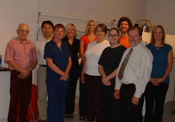
The team members in the Magnetic Resonance Imaging (MRI) and Spectroscopy (MRSI) Core Facility are, from left, Paul Knoll, Yutong Liu, Ph.D., Melissa Mellon, Lindsay Rice, Jennifer Bradley, Marie Witthoft, Erin McIntyre, Mariano Ubertei, Michael Boska, Ph.D., and Jennifer Sullivan. |
Facility name: Magnetic Resonance Imaging (MRI) and Spectroscopy (MRSI) Core Facility
Location: Department of radiology, University Tower 3, first floor
Services offered: The bioimaging laboratory scientists are available for consultation on research study design and implementation. Services range from design and performance of collaborative scientific studies with participation of facility scientists in grant and manuscript development to fee for service imaging and image processing services. Image and spectroscopic post-processing can be done either by the investigator using available laboratory computers and programs or by our in-house scientific and technical staff depending on the needs and funding levels of the investigator. Advanced scientific studies requiring custom programming and development of new imaging and image processing methods are within the scope of current activities.
Advanced MRI and MRSI techniques in current use include a wide variety of non-invasive morphological, physiological and biochemical measures of small animal (rat and mouse) tissue. Imaging methods include quantitative relaxometry (T1, T2, T2* mapping in image format), quantitative diffusion mapping, diffusion tensor imaging, quantitative magnetization transfer imaging, sensitive to demyelination of white matter tracts and high sensitivity detection of “CEST” contrast agents, vascular permeability quantification using contrast agents, quantitative (spin tagged) tissue perfusion imaging, magnetic labeling of cells for tracking cellular distribution and kinetics, and spectroscopic imaging of 1H and 31P metabolites to track cellular metabolic response to disease and therapy.
Featured equipment: Two animal MRI/MRSI systems include a Bruker Avance magnetic resonance imaging (MRI) and spectroscopy (MRS) system and a Bruker Pharmascan (7T/16 cm) MRI/MRS system equipped with Resonance Research gradients and shims. The facility includes an image processing laboratory consisting of 12 Unix workstations and two Linux (Paravision) workstations for offline data analysis and pulse program development.
Software programs available allow development of efficient processing pipelines tailored to the needs of individual projects. Custom programming methods also have been developed and implemented using Matlab, IDL and “C” on both PC and Sun workstations.
Testimonials: The core facility serves fourteen different labs on the UNMC campus and external labs from around the nation. In the past year, the lab has performed more than 650 scans including scans of mice to detect neurodegenration due to HIV infection, Parkinson’s disease and Alzheimer’s disease. In addition, studies to detect viral inflammation of the spinal cord, measure efficacy and response kinetics of nanoparticle-based therapy on drug resistant tumor cell lines and measures of the biodistribution of nanoparticle-based contrast agents for detection of rheumatoid arthritis have been performed.
Manager contact information: Michael Boska, Ph.D., at 559-3138 or mboska@unmc.edu.
Core facility managers wishing to have a focus piece done on their facilities can send an e-mail to today@unmc.edu with the facility’s name, location, services offered, featured equipment, testimonials, manager contact information and a photo of the facility.