 |
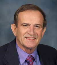
Warren Sanger, Ph.D. |
The research was reported online in the scientific journal Nature on Nov. 14 and was published in the Nov. 21 edition of the journal. Researchers at Oregon Health & Science University’s Oregon National Primate Research Center were the lead researchers on the project.
In an accompanying article in Nature News published at the same time as the Nature article, Robert Lanza, M.D., chief scientific officer for Advanced Cell Technology in Los Angeles, a leading biotechnology company, said this research breakthrough is “like breaking the sound barrier.”
Prior to the OHSU team’s recent success in a species closely related to humans, scientists worldwide have isolated stem cells only in mice using a technique called somatic cell nuclear transfer (SCNT). The method involves transplanting the nucleus of the cell, containing an individual’s DNA, to an egg cell that has had its genetic material removed. For various reasons and despite numerous attempts, previous efforts to use the SCNT technology to clone stem cells in primates have failed repeatedly.
Dr. Sanger, professor and director of the Human Genetics Laboratory at the Munroe-Meyer Institute, did genetic screening on the embryonic stem cells over the past two years to make sure that the cells contained no chromosomal abnormalities. If the cells were abnormal, they would be unsuitable for transplantation.
“This is the only stem cell project of this type we’ve ever done,” Dr. Sanger said. “It’s a real testimony to our lab staff and capabilities that we were asked to participate in the study. Only a very few labs around the country have the experience required to do this sort of testing.
“A couple other labs attempted this and were unsuccessful. It takes patience and an understanding of cell biology and genetics, which can only be achieved by experience. Our staff devoted between 300 to 400 hours on the project.”
Dr. Sanger, who was a co-author on the Nature paper, said working with embryonic stem cells involves different culture and specimen handling techniques than are used in standard genetic testing.
Prior to this project, Dr. Sanger’s lab has been involved in several research projects over the years analyzing the chromosomes of a variety of animals, including tigers, gazelles and gorillas.
“Twenty-first century science is big and complex, and major advances frequently are the result of the collaboration among elite institutions and star researchers from around the world,” said Tom Rosenquist, Ph.D., vice chancellor for research for UNMC. “This project puts UNMC scientists right at the center of one of the major advances in stem cell research, demonstrating that adult skin cells from one of mankind’s closest relatives can be the source of stem cells.
“This is the next wave of development, beyond mice and toward therapy for humans with diseases that are incurable today. The UNMC research community is proud of Dr. Sanger and his research team, who were recruited into the project because they have been recognized for decades as among the best in the world in human cytogenetics.”
Shoukhrat Mitalipov, Ph.D., director of the OHSU-based research team, was the lead scientist on the project. Don Wolf, Ph.D. of the OHSU primate center also played a significant role in the research.
“Many scientists believe that embryonic stem cells hold great promise for treating a variety of diseases including Parkinson’s disease, multiple sclerosis, cardiac disease and spinal cord injuries,” Dr. Mitalipov said. “Using our advanced methods, it is conceivable that years from now, patients could receive therapeutic embryonic stem cells developed from their very own cells meaning that there would be no concerns about transplant rejection. Another noteworthy aspect of this research is that it does not involve the use of fertilized embryos, a topic which has been the source of a significant ethical debate in this country. ”
In addition to UNMC’s Munroe-Meyer Institute, the Whitehead Institute for Biomedical Research in Cambridge, Mass., also collaborated with OHSU to conduct this research. The studies were funded by the Oregon National Primate Research Center, the National Center for Research Resources, and the National Institute of Neurological Disorders and Stroke, both components of the National Institutes of Health.
“This advance at the Oregon National Primate Research Center builds on studies supported over several years by the NCRR aimed at understanding the basic biology of stem cells and at developing methods to investigate non human primate models of disease,” said John D. Harding, Ph.D., NCRR’s director of primate resources. “These studies have great potential to accelerate progress in the field of regenerative medicine.”
The reason why Dr. Mitalipov’s team was successful when so many other previous attempts were not, lies in the method for identifying and extracting the nuclei of the eggs being used. Prior attempts resulted in damaged eggs due to the difficulties involved in removing the nucleus. This means that the eggs were not fully functional and failed to divide and develop.
To conduct the research, researchers obtained skin cells from a nine-year-old male rhesus macaque monkey at the Oregon National Primate Research Center. The researchers then used specialized imaging software called, Oosight Spindle Imaging System, to spot and remove the nuclear material attached to the egg’s spindle fibers.
The nuclei of skin cells were then inserted into nucleus-free eggs. Using this technique, two embryonic stem cell lines — groups of cells that can grow indefinitely and differentiate into any cells of the body — were successfully developed. The genetic material (DNA) of cell lines was then matched to DNA from the male donor male monkey to ensure that they were a direct clone.
Successful development of the cell lines required numerous attempts. Overall, 304 monkey eggs (oocytes) from 14 female rhesus monkeys were used to generate the two embryonic stem cell lines, a .7 percent success rate
“While development of the stem cell lines required hundreds of attempts, this research proves it can be done and will likely lead to refinements, which will make the process more efficient and lead to a higher success rate,” Dr. Mitalipov said. “This is the next step for our research team as other scientists continue to investigate the promise of stem cell therapies.”