 |
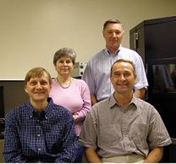
The team members of the Nanoimaging Core Facility are, first row from left, Michael Karymov, Ph.D., Alex Lushnikov, Ph.D.; second row, Luda Shlyakhtenko, Ph.D., and Yuri Lyubchenko, Ph.D. |
Core facilities, sometimes called shared resources, provide sophisticated, expensive technology and expertise that no single lab would have the financial resources to purchase and learn. These fee-for-service resources help investigators maximize productivity by providing access to technologies, services and scientific consultation at a reasonable cost. Core facilities also provide stability, reliability, cost-effectiveness and quality control that would be difficult to achieve otherwise.
Facility name: Nanoimaging Core Facility
Location: College of Pharmacy, Rooms 1019, 1019 A and 1016.
Services offered:
- High-resolution imaging of biological samples in air;
- High-resolution imaging of biological samples in liquid;
- Time lapse experiments that follow the real time changes of the sample morphology or structure;
- Nanoprobing of a sample to measure the interaction of forces between molecules;
- Study of local mechanical properties of the sample (adhesion, stiffness); and
- Data analysis for determination of homogeneity of a sample, size distribution, positioning, surface morphology, length and volume measurements.
Featured equipment:
- An integrated Atomic Force Microscope (AFM)/ optical microscope instrument;
- Stand alone MM AFM system with Nano V controller (POE); and
- The high-resolution optical system with the single molecule tracking capabilities.
The integrated AFM /inverted Olympus IX71 optical microscope is the key instrument. It is composed of a state-of-the-art AFM microscope capable imaging and probing the samples with a broad range of sizes spanning from single molecules to live cells. The AFM module of the system supports all AFM capabilities including imaging in air and aqueous solutions and equipped with the module allowing analysis of live cells. The AFM module is capable of the parallel and simultaneous analysis and probing of the same sample with both imaging modules of the integrated system. The selected Olympus IX71 inverted microscope is equipped with the module for the objective-through total internal reflection fluorescence analysis.
The stand-alone AFM (MM AFM from Veeco) is capable of high-resolution imaging of various biological samples in air and aqueous solution.
The high-resolution optical system is a new instrument that will arrive soon. The key component of this system will be a high speed and high resolution C9100-12 EM CCD camera that allows the single molecule fluorescence detection with the millisecond temporal resolution.
The facility is equipped with the off-line data analysis workstations loaded with the software required for analysis of the AFM and optical microscopy data.
Testimonials: The core facility serves several laboratories at UNMC and off-campus locations such as Mount Sinai Hospital in New York City, the University of Kansas, the University of Texas, Eastern Virginia Medical School, the University of Rhode Island and others.
Manager contact information: For more information, contact Yuri Lyubchenko, Ph.D., at 559-1971 or ylyubchenko@unmc.edu; or Luda Shlyakhtenko, Ph.D., at 559-1974 or lshlyakhtenko@unmc.edu. More details about the lab can be found online at http://www.unmc.edu/dept/pharmacy/nanoimaging/index.cfm?L1_ID=1&CONREF=1.
Core facility managers wishing to have a focus piece done on their facilities can send an e-mail to today@unmc.edu with the facility’s name, location, services offered, featured equipment, testimonials, manager contact information and a photo of the facility.