 |
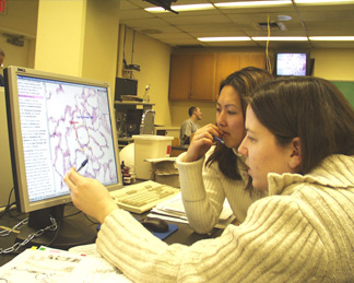
UNMC students Melissa St. Germain, foreground, and Ann Ngo study histology in a virtual microscopy lab. |
In the past, Ann Ngo and Melissa St. Germain would move slides of cellular structures under a microscope. Now, the first-year medical students are studying virtual images using a computer screen.
Professors in UNMC’s department of genetics, cell biology and anatomy are exploring the possibility of replacing aging student microscopes and slide collections with flat computer screens which allow students to view cell structures with the click of a mouse.
Virtual labs are becoming increasingly common in medical schools across the country, said Gordon Todd, Ph.D., associate professor, genetics, cell biology and anatomy, who is leading a pilot project in Wittson Hall, Room 3020, to test the feasibility and limitations of developing such a lab at UNMC.
Officials hope to obtain a grant to develop a permanent virtual microscopy lab, which would be used for a variety of courses including histology and pathology.
For the past several weeks, students, in groups of two or three, have clustered around a computer screen, while Dr. Todd projects a virtual slide overhead on a dozen classroom monitors. With the click of a mouse, students find the same image on their computers and scan, magnify and review the structure.
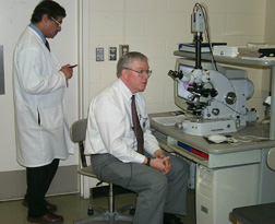 |
Shantaram Joshi, Ph.D., left, and Gordon Todd, Ph.D., work with students in the histology lab. |
Unlike the microscopes, Ngo and St. Germain recently studied a magnified view of the epithelium, complete with blue, red, yellow and green annotations, which highlighted particular areas within the respiratory system.
Students are accessing and viewing virtual slides from the University of Iowa, rather than digging through UNMC’s aging slide collection, Dr. Todd said. The technology allows everyone to see the same digitized image, Ngo said, compared with glass slides where images can vary from slide to slide. In some cases, she said, cover slips are coming off the glass slides rendering them unusable.
The virtual lab would solve an inevitable problem of replacing roughly 70 student microscopes at a cost of up to $5,000 each, as well as the expense of reproducing glass slide collections for 120 histology students, Dr. Todd said.
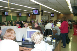 |
Histology students fill Wittson Hall, Room 3020, where faculty are leading a pilot project for a virtual microscopy lab. |
Researchers also will find the virtual lab useful because it allows them to analyze particular images with investigators across the country. Plus, the students said, the virtual lab allows them to study in places other than the library or lab.
Students can quickly learn to master the traditional microscope, if they need to, Dr. Todd said. “It’s more difficult to learn what you’re looking for in the cell structure than to learn how to use the microscope,” he said.