 |
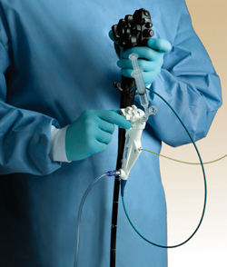
The SpyGlass camera gives doctors a view inside the bile ducts that they have never really had until now. The camera is about the thickness of a human hair. |
With the SpyGlass Direct Visualization System, gallstone removal takes less than an hour and requires no incisions. SpyGlass is a small flexible tube with a light and a video camera at the end. The new technology is able to enter tiny areas such as bile ducts that larger scopes can’t.
In the procedure, Ed Schafer, M.D., and Grant Hutchins, M.D., gastroenterologists at UNMC and its hospital partner, The Nebraska Medical Center, insert the endoscope down the patient’s throat.
When properly situated in the bile duct, the doctors use a method called lithotripsy — stepping on a pedal to send sound waves through a small wire, obliterating the gallstone that was lodged in a bile duct. The pieces of the gallstone can then be removed endoscopically.
“Before SpyGlass, oftentimes surgery would be the only option,” Dr. Schafer said. “Now with the aid of SpyGlass, the stones can be broken up within the duct and removed avoiding the necessity of surgery.”
The Nebraska Medical Center is the only hospital in Omaha with the new technology.
“This is definitely cutting-edge technology,” Dr. Schafer said. “The technology is altering the way we diagnose and treat patients with liver, gallbladder and bile duct conditions.”
“SpyGlass will provide many benefits to patients,” Dr. Hutchins added. “It will provide a better and quicker diagnosis and also cut down on the ordering of unnecessary and burdensome testing.”
Tool Improves Visualization
Another advantage of the new tool is the increased visualization of the bile ducts. The tiny SpyGlass camera — not much bigger than the thickness of a human hair — gives doctors a view inside the bile ducts that they have never really had until now.
A fiber optic probe attaches to a camera head and is inserted through a catheter that can be steered in four directions. This is designed to allow physicians to access and inspect all four quadrants of an entire treatment area. As a result, physicians are able to achieve a more accurate diagnosis for patients.
|
|
SpyGlass also is aiding doctors in the diagnosis of tumors in the pancreas and or bile ducts.
“The direct visualization allows us to discern the nature of indeterminate strictures,” Dr. Schafer said. “Then, we can directly biopsy them to determine if they are benign or malignant.”
“For cancer patients, we can biopsy with more certainty and make a more accurate plan for treatment,” Dr. Hutchins said. “Other biopsy methods created the need for additional testing or repeat procedures.”
In the tight quarters of the bile duct, the camera improves visualization markedly. If there is a concern about cancer, a good view is critical.
“If you’re off by three or four millimeters, which doesn’t sound like a lot, that’s the difference between a biopsy of cancer and normal tissue,” Dr. Schafer said. “The camera pinpoints the tumor and the tiny forceps weave up alongside to snip a small sample of the tissue.
The SpyGlass system was developed by Boston Scientific Corporation.