 |
The magnetoencephalograph (MEG) scanner — the world’s most advanced tool for studying brain function — in now at UNMC and The Nebraska Medical Center. |
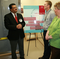 |
Sanjay Singh, M.D., associate professor of neurological sciences at UNMC and director of the MEG Center at The Nebraska Medical Center, describes the MEG’s functions during an open house at the center on Tuesday. |
The magnetoencephalograph (MEG) scanner will provide doctors the ability to look inside the brain in a way that’s never been possible before, giving patients in need of brain surgery the assurance of a safer and more effective surgery and allowing doctors to identify the region of the brain giving rise to epileptic seizures.
The Nebraska Medical Center is one of only about a dozen clinical sites in the country to have a MEG scanner.
“This new technology detects brain activity in much greater detail and with more accuracy than previous testing methods,” said Sanjay Singh, M.D., associate professor in the department of neurological sciences at UNMC and director of the MEG Center at The Nebraska Medical Center.
This kind of view into the brain will give doctors the ability to identify where the brain is malfunctioning with increased precision, it will provide neurosurgeons greater precision when operating and it will give epileptologists an increased ability to identify where the brain is malfunctioning.
“MEG is potentially the most helpful tool for treating patients with neurological illnesses like epilepsy,” said Dr. Singh, who also is director of the Nebraska Epilepsy Center.
MEG will also allow doctors to explore and gain a better understanding of other neurological and psychiatric diseases like depression, schizophrenia, autism, dementia, Alzheimer’s disease and Parkinson’s disease.
“Unlike other testing methods, the MEG scanner gives us the ability to look at the brain in real time,” Dr. Singh said. “For example, we get to look inside the brain when a person is solving complex problems like mathematics, when a person is feeling happy or sad and when a person responds to a noise.
“If a person moved his finger, the MEG would also allow us to see what part of the brain told the finger to move.”
The MEG works by measuring and recording the brain’s magnetic fields that are created by the brain’s electrical activity. The scanner detects instantaneous changes in brain activity, allowing doctors to track changes that happen in milliseconds
“During a MEG scan, we get to see the brain in action,” said Tony Wilson, Ph.D., MEG scientist and assistant professor in the department of neurological sciences. “During the MEG scan, we may ask a patient to read a specific word, and we can see the word appear in the visual areas of the brain first, then get transferred to the language areas of the brain, and then to the speech areas.
“The MEG is the future,” Dr. Wilson said. “Compared with other brain imaging technologies, it has the unique ability to monitor brain ‘dynamics.’ MEG takes pictures with millisecond precision, therefore, I can see brain areas become active and communicate with other brain areas to produce complicated actions and thoughts.”
|
|
“One of the great challenges of brain surgery is removing the diseased part of the brain, while sparing healthy brain tissue that controls vital functions,” said Deepak Madhavan, M.D., assistant professor in the department of neurological sciences at UNMC and associate director of the MEG Center. “The MEG will give neurosurgeons a road map of the brain to help them identify the areas to be removed and help to preserve the critical areas of the brain.
“For a patient with epilepsy who has had no luck controlling epilepsy with medications, the MEG opens up a whole range of surgical options to becoming seizure free. The MEG allows us to localize the area of seizure generation or the seizure onset area with greater precision so that neurosurgeons can perform a more complete surgical removal of that area.”
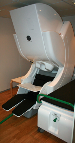
The non-invasive, outpatient MEG scan takes about an hour. MEG recordings are done in a special shielded room that blocks magnetic fields in the environment. This ensures that only the magnetic fields generated by the brain are detected by the MEG sensors. The patient sits underneath a helmet shaped area, which resembles a 1950s salon hair dryer. The helmet has 306 super-cooled sensors that measure changing patterns of the brain’s magnetic activity.
After the MEG scan, the MEG recordings are combined with a magnetic resonance imaging (MRI) scan, which shows the actual structure of the brain. The combined scan, called magnetic source imaging (MSI), shows areas of the brain that may generate seizures as well as localizes areas of normal brain function with precise timing. MSI can provide neurosurgeons a detailed “map” of the brain.
Joint funding for the MEG scan was secured from an anonymous donor by the Office of Development at The Nebraska Medical Center and the medical center funded the remainder of the expense.
“The MEG technology is the best thing that has happened to neurosciences in a long time,” Dr. Singh said. “The clinical uses of the MEG are many and the research possibilities are endless.”