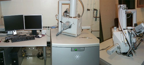
Serial Block Face Scanning Electron Microscope (SBF-SEM)
In May 2019, the EMCF was awarded an NIH S10 shared instrumentation grant (1S10OD026790-01) for the purchase of a Thermo Scientific Volume Scope. This is a field emmision SEM with an in-chamber ultramicrotome that allows for serial sectioning of a sample embedded in epoxy resin. After each section, the microscope takes an image of pre-defined ROI's which results in a stack of images through the sample at a z-resolution down to 40nm. This can be pushed further to a z-resolution of 10nm with the use of higher beam energies followed by multi-energy deconvolution (MED). The sample must be ideal in order for this technique to be successful. Raw images from the microscope are then aligned and sub-cellular features can be manually segmented out using the Amira software. This results in a 3D model of the sample that can reveal cellular features and relationships that could only be discerned on the TEM after months of work, if ever. The ultramicrotome can also be removed and the microscope can be operated in high resolution mode for standard SEM imaging. The microscope also features "low vacuum" mode which uses an atmosphere of water vapor in order to mitigate charging on non-conductive samples.
TEM
The EMCF installed a new FEI Tecnai G2 Spirit transmission electron microscope in 2009 thanks to the support of the NIH Shared Instrument grant (NIH 1 S10 RR024650 01A1). The instrument is a 120kV, LaB6 equipped microscope and has a goniometer stage and tomography holder for precise tilting of specimens for stereoscopic images, or to orient specimens for optimal viewing. Images are acquired digitally with an AMT digital imaging system.
SEM
For scanning electron microscopy, the facility is equipped with a FEI Quanta 200 SEM installed in 2006. Funding for this instrument was provided by the Office of the Vice Chancellor for Research as part of the Nebraska Tobacco Biomedical Research & Development Fund. The Quanta can operate in conventional high vacuum mode, variable pressure low vacuum and environmental SEM modes. This range of modes allows for examining non-conductive, beam sensitive and wet specimens as well as conventionally prepared SEM samples. The instrument is also equipped with a new Bruker AXS Quantax XFlash 4010 x-ray microanalysis detector, allowing for elemental microanalysis of SEM samples. The most recent upgrades to the SEM included a beam deceleration module, which allows for better surface detail to be acquired from beam-sensitive samples. In addition, a NavCam accessory was added in 2015, which assists users in locating regions of interest from multiple samples. All images are acquired digitally.
Support Equipment
The facility has supporting equipment for all standard biological specimen preparation protocols.
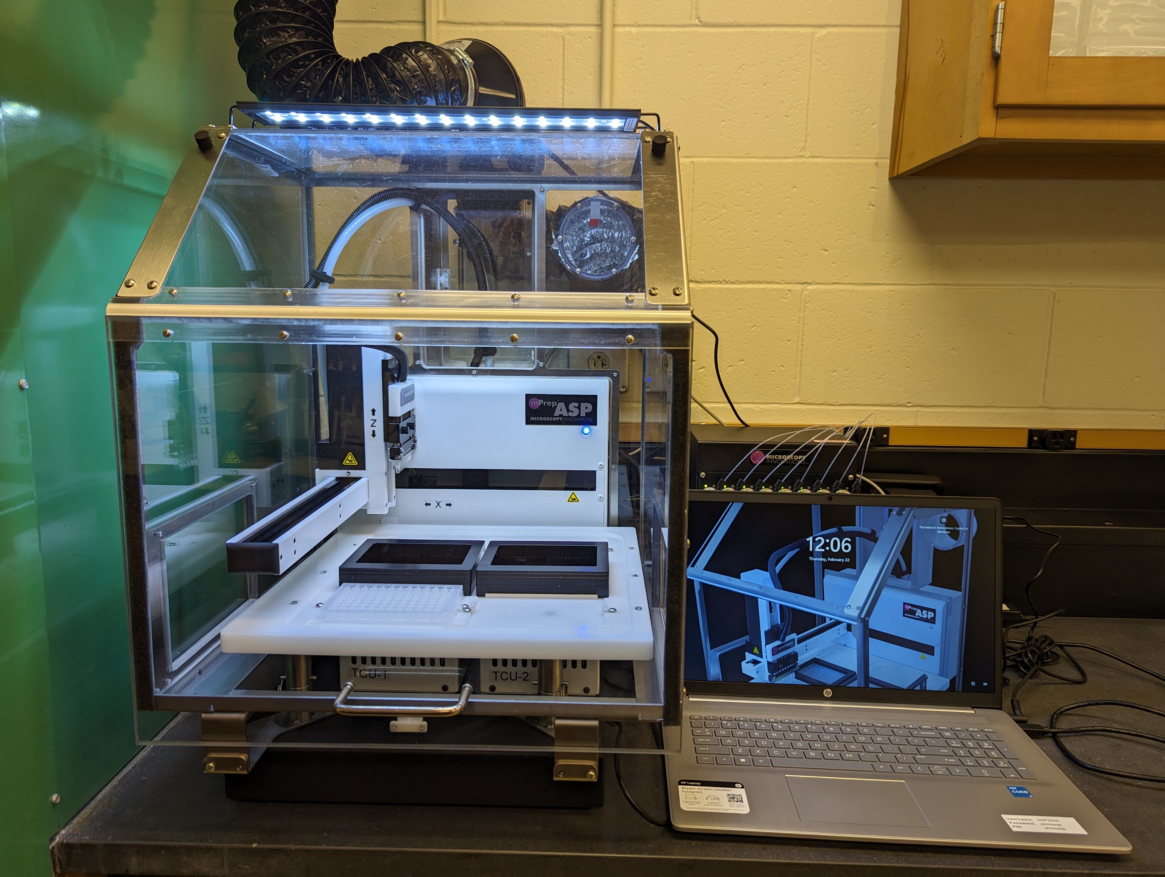
Automated Sample Processing
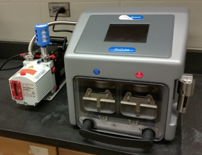
New! In June 2023 the EMCF acquired an ASP-2000 automated sample processing unit from Microscopy Innovations. Support for this equipment came from the VCR Cores and the Nebraska Research Initiative (NRI). The instrument features an 8-channel pipette head with the ability to stack 2 sample tubes for a total capacity of 16 samples per run. It uses constant agitation and advanced fluid flow dynamics to reduce processing time from 8 hours to approximately 4. The ASP-2000 features 2 temperature control plates that allow for precise incubation temperatures depending on the needs of the protocol. The protocols are easily modified and shared with other ASP users. Because of the temperature control units (TCUs), this instrument is fully capatible with serial block face imaging sample prep protocols, taking a 3 day protocol and condensing it to approximately 8 hours. Samples can be taken from their fixed state all the way through epoxy resin embedding with minimal intervention from lab staff. This instrument greatly improves sample turn around time and lends itself both to basic processing as well as more experimental techniques such as immunogold labeling.
TEM
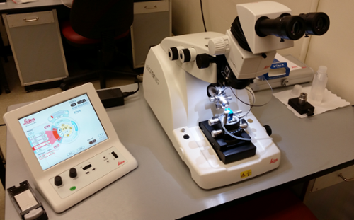
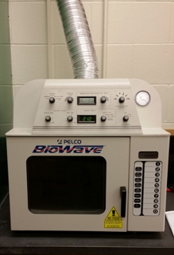
This includes a Leica Ultracut ultramicrotome, LKB glass knifemakers, diamond knives, centrifuges, and embedding oven. A Pelco UV-2 cryochamber is available for low temperature embedding for immunocytochemistry and a Pelco Biowave variable wattage microwave fixation system is used for expedited protocols and increased solution penetration. In addition, a GloQube glow discharge system from Quorum Technologies was added in November, 2016 thanks to funding by the Nebraska Bankers Association. This system allows TEM grids to be plasma treated in order to impart various surface characteristics (hydrophilic, hydrophobic, negative charge, etc.).
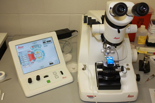
A Leica Ultracut UC7 was added in June, 2017 thanks to funds from the Nebraska Research Initiative.
SEM
Support equipment for SEM includes a Pelco critical point drying apparatus and a Hummer VI sputter coater.