This page outlines the various equipment and resources available within the MITI Core Facility space, including:
- General Equipment Information
- Neurotar Mobile Home Cage
- Olympus FVMPE-RS advanced multiphoton microscope
- Spectra-Physics InSight X3 dual-line, tunable multiphoton laser for ultrafast, multichannel imaging
General Equipment
- Insight tunable laser from 680-1300nm w/ fixed 1045nm laser
- 458nm and 588nm stimulation (only) laser lines
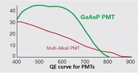
- 2 multi-alkaline detectors, 2 GaAsP detectors
 Filters from Violet to Cy5
- FV30-FVG: EM: 410-455nm, 495-540 nm
- FV30-FCY: EM: 460-500, 520-560 nm
- FV30-FGR: EM: 495-540nm, 575-645 nm
- FV30-FCY: EM 575-645nm, 660-750 nm
- Objectives
- 5x air objective, 0.15 NA, 20mm WD (live or fixed samples)
- 25x water immersion, intravital objective, 1.05 NA, 2mm WD (live samples)
- 25x multi-immersion objective, 1.0 NA, 4mm WD (optically cleared samples)
- Ideal for volumetric imaging of small, optically cleared samples.
- For volumetric imaging of large, optically cleared samples, see affiliated AMCF equipment.
- Stage heater for extended intravital imaging
- Available isoflurane system
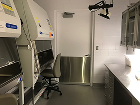
- Attached room is available for sample preparation
Neurotar Mobile Home Cage
The Neurotar Mobile Home Cage integrates real-time imaging with behavioral/locomotion tracking.
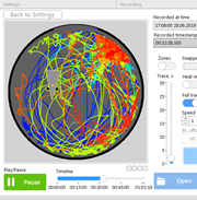
Perform real-time imaging of neuronal activity (brain, spinal cord) concurrently with activity tracking using this system. To utilize this system, head plates (model 1-11, shown below) and other imaging accessories may be ordered through Neurotar.
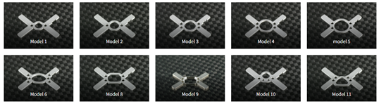
Custom 3D printed adaptors can also be used, with this stabilization arm being used to stabilize other types imaging windows (dorsal skin fold, abdominal). Custom 3D printed designs may be discussed with Dr. Jensen-Smith.
Locomotion tracking software is available for download, and other publications and educational webinars for the mobile home cage are available on the Neurotar website.
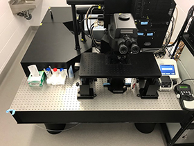
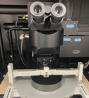
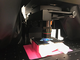
Olympus FVMPE-RS Multiphoton Microscope
The Olympus FVMPE-RS Multiphoton Microscope is a multichannel, multiphoton excitation system specifically tailored for intravital and deep tissue imaging. It delivers high speed (up to millisecond imaging) for rapid in vivo imaging of physiological responses, near infrared excitation for deep tissue imaging. Our system is equipped with both a high-resolution galvanometer-based scanner for deep tissue imaging and the fastest resonant scanner on the market for ultrafast imaging.
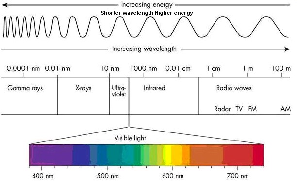
The dual laser, multiphoton system and detectors allow simultaneous image capture of up-to four different fluorophores (blue (410-455 nm), green (495-540 nm), short reds (575-645 nm) and long reds (660-750 nm) with detection capacities extending to 850nm.
GaAsP PMT and Multi-Alkali PMT detector response profiles:
Additional information available on the Olympus website.
By implanting an imaging window chamber (dorsal, abdominal or brain), real-time studies of various biological processes (cell migration, drug delivery and clearance, tumor grow, etc..) may be imaged for weeks to months. We may be able to generate custom imaging windows for your specific research needs, please contact Dr. Jensen Smith to discuss and determine feasibility (price varies by complexity and composition).
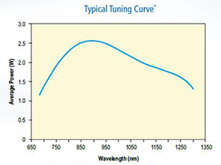
Spectra-Physics InSight X3 dual laser system
The Spectra-Physics InSight X3 dual laser system features a broad 680 nm to 1300 nm tunable laser with high average and peak power levels across the tuning range. The dual beam option on our system also contains a fixed 1040 nm laser line. This synchronized, tunable, ultrafast (80 MHz), two output beam system allows simultaneous imaging of various fluorophores including; GFP and mCherry, as well as, collagen second and third harmonic generation (SHG/THG) imaging. The DeepSee dispersion pre-compensator optimally delivers short pulses (< 120 fs) to samples allowing maximum fluorescence and penetration depth.
Training and access to the imaging system, support in the design of experimental procedures, acquisition of image data, and analysis of the required images are offered through the MITI Core. Services are available to investigators within the University of Nebraska network, as well as, academic or commercial/industrial institutions in the region.
Please ensure compliance and discuss with Dr. Jensen Smith all intramural and extramural regulations regarding use of research animals and hazardous materials before using the system. Contributions made by the core facility should be briefly mentioned in the acknowledgments of scientific publications. If the work performed in the core facility is more than routine, publication as co-author is appropriate.